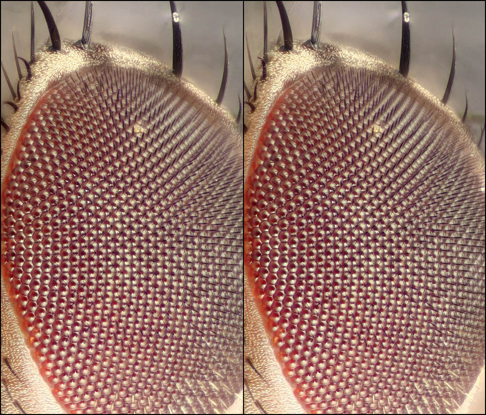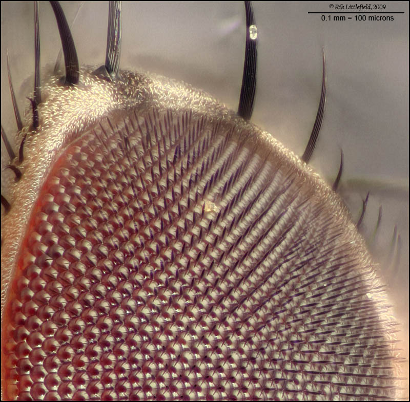
I thought I'd try an eye with setae, just for comparison.
Compare this with the naked fly's eye at http://www.photomacrography.net/forum/v ... php?t=7373.
This is a fruit fly, about 1/2 frame.
Canon 300D camera, Olympus CH microscope base, Nikon CF M-Plan 20X NA 0.40 ELWD objective at 21X, pingpong ball diffuser with dual fiber halogen illumination, 0.5 second at ISO 100, 106-frame stack at 4 microns focus step, Zerene Stacker PMax, no retouching.
It's interesting to notice, in this picture, the ommatidia at lower left. They appear to be very complicated. But in fact, they are just smooth shiny bumps --- it is their environment that is very complicated! In each ommatidium, the dark disk is a reflection of the microscope objective, while the lighter ring is a reflection of the illuminated pingpong ball. The radial rays are simply reflections of the surrounding setae, of which there is one at almost every junction between ommatidia. One of the advantages of electron microscopy, such as shown at http://commons.wikimedia.org/wiki/File: ... d_eye_.jpg, is that there are no confusing reflections.
--Rik
Edit: to provide a new link to a scanning electron image, since the previous one disappeared.