I remember a conversation I once had with Ron Neumeyer a few years back about the productivity of one of his favorite sampling spots, and particularly whether he had found anything interesting. "The usual suspects" was his reply.
For me, one of the most intriguing aspects of microscopy is the excitement of seeing something for the first time. Sometimes a slide is loaded with subjects, but the are all familiar "faces".
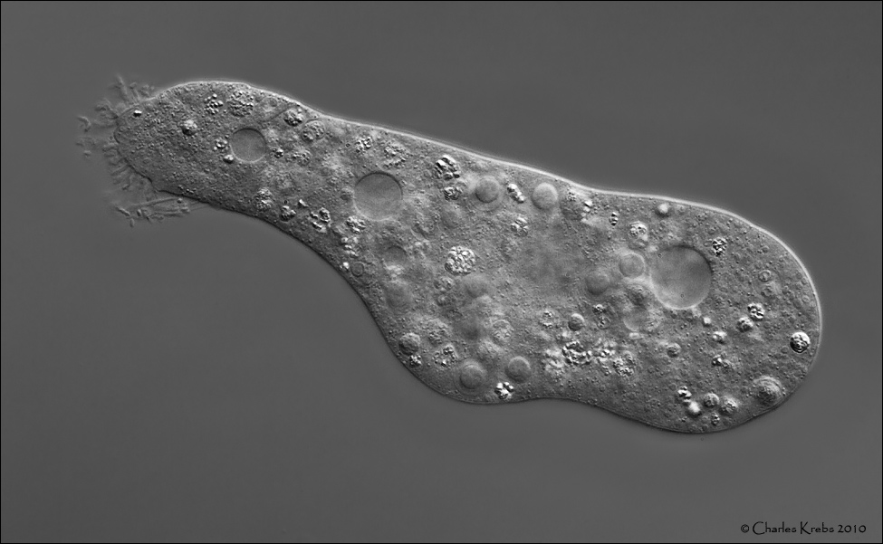
But it doesn't stop me from taking a few pictures ... so here are a few of the "usual suspects" from a slide I looked at last night.
Olympus BHS. Olympus 60/1.40 S Plan Apo. Olympus NFK 1.67x photoeyepiece.DIC illumination with electronic flash. Canon 50D.
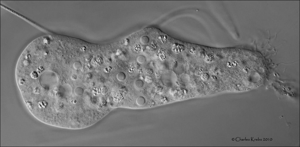
Olympus BHS. Olympus 60/1.40 S Plan Apo. Olympus NFK 1.67x photoeyepiece.DIC illumination with electronic flash. Canon 50D,
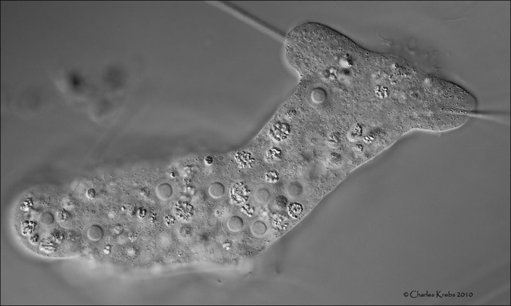
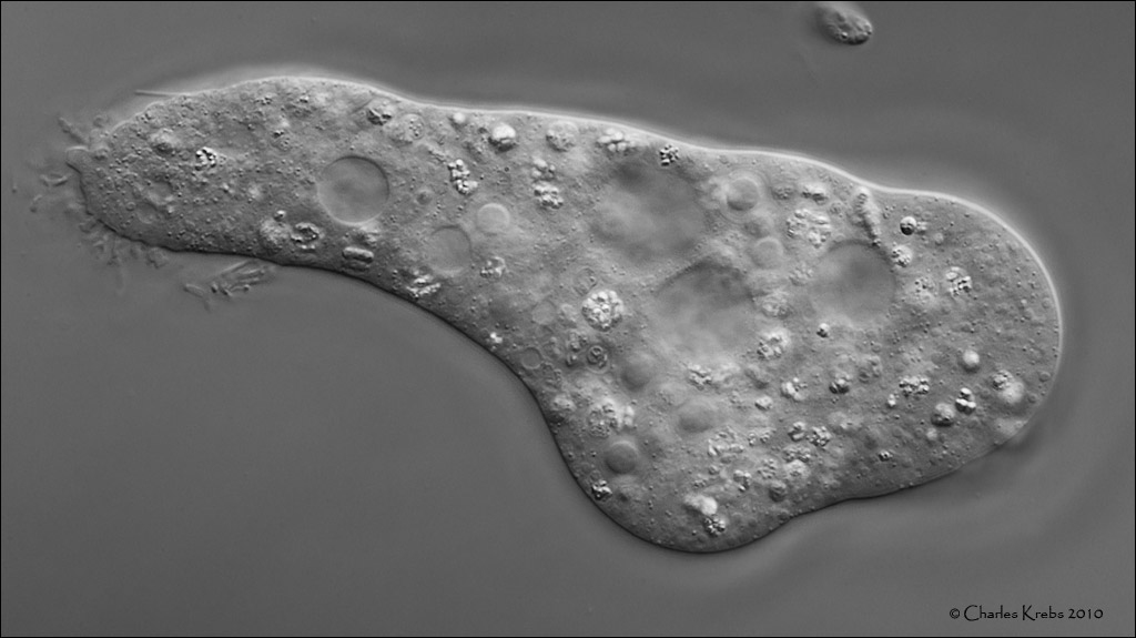
Olympus BHS. Olympus 20/0.70 S Plan Apo. Olympus NFK 1.67x photoeyepiece.DIC illumination with electronic flash. Canon 50D.
Last edited by Charles Krebs on Mon Dec 13, 2010 2:57 pm, edited 1 time in total.
Exquisite.
The amoeba looks like a Saccamoeba.
If you haven't seen the movie "The usual suspects", it's worth seeing.
NU.
student of entomology
Quote – Holmes on ‘Entomology’
” I suppose you are an entomologist ? “
” Not quite so ambitious as that, sir. I should like to put my eyes on the individual entitled to that name.
No man can be truly called an entomologist,
sir; the subject is too vast for any single human intelligence to grasp.”
Oliver Wendell Holmes, Sr
The Poet at the Breakfast Table.
Nikon camera, lenses and objectives
Olympus microscope and objectives
It seems that the amoeba has several nuclei. There are at least six visible. That makes the species-distinction very tricky. I don't know any amoeba of this type (e.g. Trichamoeba or Saccamoeba) to be multi-nucleate. Do you have more pictures of this amoeba and how large is it? It is a very interesting one.
Here are a few more of the amoeba if you want to try to figure out what it might be. Plenty of nuclei to look at here! It measures about 135 micron long, by about 43 micron wide when "configured" as in the first picture posted.
Thank you! I'm quite sure this species has never been described before. I will add it to my database and rescan my literature. If I find new data concerning this species, I let you know.
Congratulations, it is always fascinating to discover something that is new for science, to feel a little like Lewis and Clark!
"An Illustrated Guide to the Protozoa" (2nd ed) suggests that the amoeboid motion in Pelomyxa is distinctive "with the cytoplasm flowing along the center of the body and spreading out, fountain-like, at the anterior end." I wasn't quite sure what to make of the next sentence, regarding feeding. "The posterior end of the cell has a bulbous or filose uroid, and food particles may be ingested here." Are they saying it eats with its tail?
Other features are that Pelomyxa are found in anaerobic or micro-aerophilic environments and that the amoeboid form can reach 3mm long.
These sound like very interesting micro-organisms due to their complex life cycle. "In spring, cysts release small binucleate amoebae, which grow and become multinucleate and acquire endosymbiotic bacteria. They become elongate with posterior uroid, and flagella are evident at this stage. The cells later become spherical, and the endosymbiotic bacteria congregate around the nuclei. These cells fragment into rosettes giving rise either to cysts in winter or to small amoebae whch may undergo another cycle of development."
(Note: I think that the flagella being refered to are very short and difficult to observe with light microscopes.)
I'm not sure if what Charles has found fits with this description, but since I happen to have this book checked out from the library, I thought that I would share what I found in it.
Pelomyxa and Chaos are quite different from this amoeba. Both do have nuclei with fragmented nucleoli, often in the periphery of the nucleus. Pelomyxa has a lot of symbiotic bacteria and Chaos is much larger and has a locomotive form with dorsal folds.
The only amoeba which resembles this one is Gruberella flavescens, it has 1-37 spherical nuclei with a central nucleolus, but it is a marine species.
Those hairs are not cyanobacteria, but villi: thin, hairlike protrusions of the protoplasm of the amoeba. You can see them at the posterior end, which is called the uroid.
Ferry, I meant the thick round object coming out of the top left corner of the first of the last three images, and again out of the top middle of the second image. Those don't seem to be part of the amoeba.
Mitch, sorry for the misunderstanding
Well, in the second image the amoeba is flowing around the cyanobacterium, maybe to see if it is food. So the cyanobacterium comes really out the amoeba.