Paramecium bursaria
Moderators: rjlittlefield, ChrisR, Chris S., Pau
- ralfwagner
- Posts: 441
- Joined: Sat Aug 05, 2006 8:34 am
- Location: Germany, Duesseldorf
- Contact:
Paramecium bursaria
Hello,
Paramecium bursaria is for sure a classic object in amateur microscopy and often discussed here. I like to show here a small animation demonstrating the change of the two contractile vacuoles. My animation changes every 0,8 s, the real time delay betwen the pictures is 4 s. Furthermore the macronucleus and the cilia can be seen in this phasecontrast pictures.
----------------------
Ralf
http://www.lichenes.de
http://www.dr-ralf-wagner.de
http://www.youtube.com/user/drralfwagner
Ralf
http://www.lichenes.de
http://www.dr-ralf-wagner.de
http://www.youtube.com/user/drralfwagner
- ralfwagner
- Posts: 441
- Joined: Sat Aug 05, 2006 8:34 am
- Location: Germany, Duesseldorf
- Contact:
Mitch640 wrote: I'm surprised he stayed in one spot long enough for a couple of frames.
You must not be surprised! Indeed that critter moved a little bit between these two shots and I had to adjust the two pictures manually.
----------------------
Ralf
http://www.lichenes.de
http://www.dr-ralf-wagner.de
http://www.youtube.com/user/drralfwagner
Ralf
http://www.lichenes.de
http://www.dr-ralf-wagner.de
http://www.youtube.com/user/drralfwagner
-
- Posts: 156
- Joined: Sun Jan 02, 2011 11:50 am
Excellent!
Have you tried to immobilize the paramecia? Adding things like agarose would drastically increase the viscosity of the medium which will halt the critter in one place. Then you can make a consecutive set of images that show the cytoplasmic movements, contractile vacuoles and beating cilia over a wider time range.
- ralfwagner
- Posts: 441
- Joined: Sat Aug 05, 2006 8:34 am
- Location: Germany, Duesseldorf
- Contact:
bromodomain wrote:
Have you tried to immobilize the paramecia? Adding things like agarose would drastically increase the viscosity of the medium which will halt the critter in one place.
I do know this method, but didn't try it so far, although I have a bottle of 4% Methylcellulose in my lab. Nevertheless this critter was immobilzed simply by water evaporation. Indeed he exploded a few seconds after the last shot.
----------------------
Ralf
http://www.lichenes.de
http://www.dr-ralf-wagner.de
http://www.youtube.com/user/drralfwagner
Ralf
http://www.lichenes.de
http://www.dr-ralf-wagner.de
http://www.youtube.com/user/drralfwagner
-
- Posts: 156
- Joined: Sun Jan 02, 2011 11:50 am
No, it exploded from drying out. As the drop of water under the coverslip dries out, the bugs are put under duress by the lack of oxygen, rising levels of minerals being left behind and the heat of the illimination.
Single celled animals spend a very high portion of their energy just keeping their cell membrane intact. When their oganelles start failing, one of the first things to go is the cell wall. It only takes a failure of one tiny spot on the membrane to tear open, which instantly kills the animal and deflates it. It looks like a slow motion explosion.
-
- Posts: 156
- Joined: Sun Jan 02, 2011 11:50 am
I've always thought it was because the the coverslip crushed the cells. They always get really flattened and I can see every detail of their anatomy in the same focal plane. If the salt concentration rises as water evaporates should that result in efflux of water from the cell thus causing it to shrink? At least thats what happened to my blood cells when I put them in saline. Im not 100% sure but I think thats why marine protozoa dont have contractile vacuoles.
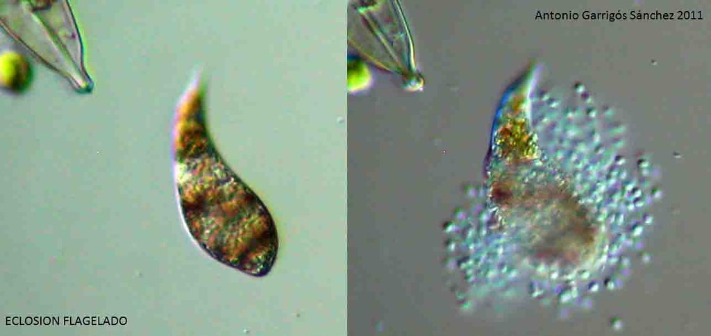
I'm just a rank amateur about microbes, but I came across an article about microbe cell walls and certain organelles in the cell dedicated to just keeping the cell wall intact. As conditions deteriorate for the cell, other organelles will shut down in favor of keeping the ones that constantly repair the cell wall, running. When they fail, the wall tears and the animal dies.
I have never run across the problem of the coverslip lowering enough to flatten a cell, since all my slides have been fitted with 4 little dots of nail polish to prevent them from crushing the bugs. One thing I don't have, is a good mental picture of the size of the bugs in relation to everything else, so I find it hard to imagine the coverslip crushing them, although it could happen. I know it does happen with bigger things like copapods. At any rate, the little microbes still explode, even under a raised coverslip.
I didn't know about marine protozoa not having contractile vacuoles either. I live a thousand miles or so inland. I learn something every day here.
- ralfwagner
- Posts: 441
- Joined: Sat Aug 05, 2006 8:34 am
- Location: Germany, Duesseldorf
- Contact:
I think so, too: crushed by pressure. When I was saying "simply by water evaporation" this was not the whole truth. First I sucked off a large amount of water with a towel. Evaporation time left was only 2 minutes. I was sucking off the water first because according to my experience only a flat critter will show us a nucleus and contractile vacuoles. And, of course, a flat critter dosn`t move that fast anymore.bromodomain wrote:So it was crushed by the pressure. I noticed this helps to get all the internal structures in the focal plane. But yeah you'll have to be quick unless you are interested in the squashed contents.
Would like to see more
Anyway, there is always something to learn here. I didn't know that marine critters do not have a contractile vacuole, too.
----------------------
Ralf
http://www.lichenes.de
http://www.dr-ralf-wagner.de
http://www.youtube.com/user/drralfwagner
Ralf
http://www.lichenes.de
http://www.dr-ralf-wagner.de
http://www.youtube.com/user/drralfwagner
Hi,
Bromodomain wrote:
"Im not 100% sure but I think thats why marine protozoa dont have contractile vacuoles."
I have often read this as well, but it is not the case. Many marine ciliates have prominent contractile vacuoles. However, in my experience they do not discharge anywhere near as frequently as freshwater ciliates. It is thought that the function in seawater may be removal of water ingested with food/ excretion of excess sodium and/or calcium / removal of other wastes (ie nobody knows!).
If you try and slow down ciliates with agarose, methyl cellulose or similar substances you always, in my experience, slow down the vacuole discharge and often with time the vacuole swells up and ceases to discharge...often a precursor to a small ciliate explosion !
A question for you Ralf...is this an animation of several shots or just cycling between two ? I´m intrigued by the apparent regular alternate discharge. Thanks for a great posting...
muito obrigado,
boa sorte,
Brian
- ralfwagner
- Posts: 441
- Joined: Sat Aug 05, 2006 8:34 am
- Location: Germany, Duesseldorf
- Contact:
BJ wrote: A question for you Ralf...is this an animation of several shots or just cycling between two ? I´m intrigued by the apparent regular alternate discharge. Thanks for a great posting...
Brian
Thank you, Brian. It is only cycling between two shots.
----------------------
Ralf
http://www.lichenes.de
http://www.dr-ralf-wagner.de
http://www.youtube.com/user/drralfwagner
Ralf
http://www.lichenes.de
http://www.dr-ralf-wagner.de
http://www.youtube.com/user/drralfwagner
- Antonio Garrigós Sánchez
- Posts: 28
- Joined: Thu May 12, 2011 10:52 am
Nice sequence Ralf.
The hatches are interesting, internal organelles are dispersed quickly by the media.
A picture of this process is not of good quality, but I think cross-hatch of a Euglena
Eclosión flagelado por Antonio Garrigós Sánchez
The hatches are interesting, internal organelles are dispersed quickly by the media.
A picture of this process is not of good quality, but I think cross-hatch of a Euglena
Eclosión flagelado por Antonio Garrigós Sánchez