Camellia pollen

Images made through a microscope. All subject types.

Moderators: rjlittlefield, ChrisR, Chris S., Pau

Charles Krebs
Posts: 5865
Joined: Tue Aug 01, 2006 8:02 pm
Location: Issaquah, WA USA
Contact:

Camellia pollen

Post by Charles Krebs »

I'll continue the pollen parade with this Camellia pollen. Tried it "dry" under cover slip, in immersion oil, and in glycerin. "Dry" looked the most natural, and showed the best surface texture. Immersion oil cleared out all the color. Glycerin worked well and did not remove the yellow color.

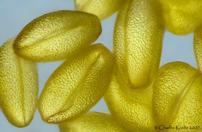
The top shot is "dry", from a stack of 22 made with the 40X objective.
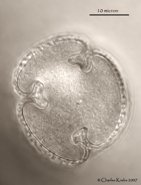
The second shot (converted to b+w) is most helpful to understand the shape clearly. It is an "optical section", a cross section of a pollen grain that is standing on one end. Taken with the 100X and looking through the end to about the midpoint, it is clear how the grooves seen in the top image continue inward and flare out into heart shaped channels.

These pollen grains averaged about 30 micron in diameter with a length of about 55-60 micron.

Image

Image

Ken Ramos
Posts: 7208
Joined: Thu Jul 27, 2006 2:12 pm
Location: lat=35.4005&lon=-81.9841

Post by Ken Ramos »

Amazing Charlie :o Seems folks are getting on the band wagon with these things :lol: Wonderful shots! :smt023

rjlittlefield
Site Admin
Posts: 23599
Joined: Tue Aug 01, 2006 8:34 am
Location: Richland, Washington State, USA
Contact:

Post by rjlittlefield »

Charlie, you've pushed the bar up another notch or two with these shots. Great stuff!

But of course, I'm confused, and I want more details. :D

I'm presuming that these are transmission DIC, backlit. It seems clear from the "optical section" that your lenses and the nature of the subject allow seeing sharp detail inside the grains. In the first pic, the image appears sharp all the way from front surface through the outer edge of at least some grains. That means (judging from the cross section) that in the first pic you must have stepped focus clear through at least one of those T structures. I would expect that when you're focused on the edge of the arms of the T, they would be visible. So......

Why don't we see even a hint of the T's -- or any other interior detail -- in the first shot?

Do the T's disappear from the composite because they are overwhelmed by higher contrast surface detail, or do the T's not show from the side even in individual frames focused on them?

Thanks! :D

--Rik

Charles Krebs
Posts: 5865
Joined: Tue Aug 01, 2006 8:02 pm
Location: Issaquah, WA USA
Contact:

Post by Charles Krebs »

Rik,
I should have mentioned that the second shot was from a slide that had the pollen in immersion oil. The immersion oil very quickly removes all the yellow coloration (it dissipates into the oil) and the grains become as clear as glass. (Makes me wonder about the location and nature of the yellow pigment... but that's another issue). With the lighting used there was much less ability to "see through" when the pollen was in air and the yellow coloration was in place.

One of the reasons I converted to b+w was that there was no more color in the pollen grain, and the oil around it had a strong variable yellow tinge.

Here's an animated gif of pollen in immersion oil. I happened to find one in profile and one on end right next to each other. They were clear and transparent, so you can see the "channel" as you focus into it.
Image

discomorphella
Posts: 607
Joined: Sun Oct 01, 2006 7:26 pm
Location: NW USA

Post by discomorphella »

Hi Charlie--
These are spectacular shots, as usual. Now I must rush out and collect some pollen. Might as well get something out of pollen besides a runny nose and itchy eyes...

--David

Ken Ramos
Posts: 7208
Joined: Thu Jul 27, 2006 2:12 pm
Location: lat=35.4005&lon=-81.9841

Post by Ken Ramos »

Cool 8)

rjlittlefield
Site Admin
Posts: 23599
Joined: Tue Aug 01, 2006 8:34 am
Location: Richland, Washington State, USA
Contact:

Post by rjlittlefield »

Charles Krebs wrote:With the lighting used there was much less ability to "see through" when the pollen was in air and the yellow coloration was in place.
Hhmm... :-k

So, if I understand correctly, when the pollen is dry, your DIC setup does not see clearly inside the grains as I had supposed, but instead just picks up surface texture.

Fascinating!

That certainly makes me wonder about the distribution and other characteristics of the yellow stuff. Seems like if you can't see detail through it, but light gets through it to illuminate the near surface, then it must be strongly diffusing. Have I missed something here? :?:

--Rik

Post Reply Previous topicNext topic