a "What Is It?" (too easy I think!)
Moderators: rjlittlefield, ChrisR, Chris S., Pau
-
Charles Krebs

- Posts: 5865
- Joined: Tue Aug 01, 2006 8:02 pm
- Location: Issaquah, WA USA
- Contact:
a "What Is It?" (too easy I think!)
Nikon MM-11 microscope, Olympus 50/0.50 LMPlanFL N , Olympus TLU tube lens, 50X on sensor, Canon T3i
-
rjlittlefield

- Site Admin
- Posts: 23608
- Joined: Tue Aug 01, 2006 8:34 am
- Location: Richland, Washington State, USA
- Contact:
-
rjlittlefield
- Site Admin
- Posts: 23608
- Joined: Tue Aug 01, 2006 8:34 am
- Location: Richland, Washington State, USA
- Contact:
By the way, this strikes me as astonishingly clean for 50X on sensor. If it weren't for the scale bar and the description, I would have no idea how small this subject is.
Very nice in all respects.
I'd ask for a stereo, but with this illumination and posing, I suspect the 3D wouldn't add much!
--Rik
Very nice in all respects.
I'd ask for a stereo, but with this illumination and posing, I suspect the 3D wouldn't add much!
--Rik
-
Charles Krebs
- Posts: 5865
- Joined: Tue Aug 01, 2006 8:02 pm
- Location: Issaquah, WA USA
- Contact:
Yes that's it of course. I had actually forgotten about the post you referenced... remembered two older ones...
http://www.photomacrography1.net/forum/ ... php?t=1997
http://www.photomacrography1.net/forum/ ... php?t=2003
... but I figured after 7 years it might stump someone
The 50/0.50 is a new one for me. I was a little leery of using it because the NA is pretty modest for a 50X (well into diffraction territory) but the 10.6mm working distance is decent. I'm piecing together a scope for this type of subject and I'm using an Olympus tube lenses and a some of their objectives (and a 5X and 10X Mitutoyo). The new Olympus UIS2 FL (fluorite) objectives look quite good.
-
Charles Krebs
- Posts: 5865
- Joined: Tue Aug 01, 2006 8:02 pm
- Location: Issaquah, WA USA
- Contact:
I suppose I should describe what this is here, rather than just providing older links...
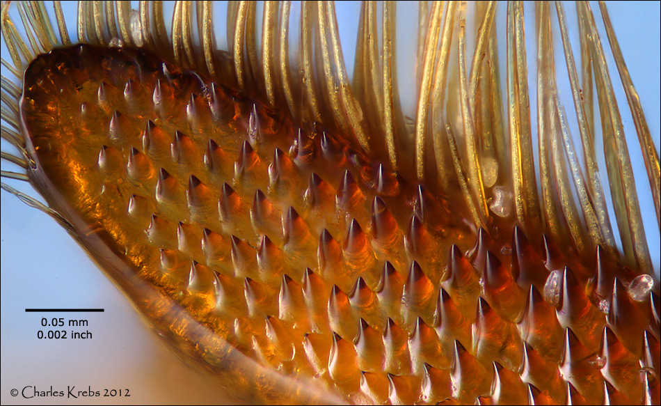
This is the top of the tarsus on a rear leg of a honeybee (at the joint where it meets the tibia... the large joint in the honeybee's rear leg). The area where these leg segments meet is known as the "pollen press" or "pollen packer". Pollen is moved into this area via stiff hairs or "combs" on the bees legs, and when these two rear leg segments are "flexed" the pollen is compressed (like a "nutcracker" action). It is then moved and collected on the outer surface of the tibia... the "pollen basket".
I tried to clear out all pollen so the interesting structure would be visible, but you can still see a few blackberry pollen grains.