Spirostomum & ???

Images made through a microscope. All subject types.

Moderators: rjlittlefield, ChrisR, Chris S., Pau

RogelioMoreno
Posts: 2979
Joined: Fri Nov 20, 2009 11:24 am
Location: Panama

Spirostomum & ???

Post by RogelioMoreno »

Here a picture of Spirostomum and a set of pictures of a ciliate that I would like to know the ID (maybe Chilodonella?).

Spirostomum
Image

The following set of ???
Image

Look at the detail inside the mouth
Image

Image

Image

Rogelio

Wim van Egmond
Posts: 826
Joined: Tue Aug 15, 2006 9:28 am
Location: Berkel en Rodenrijs, the Netherlands
Contact:

Post by Wim van Egmond »

I am not sure about Chilodonella. They ave a different shape. Perhaps a type of Nassula?

This contrast rich deep polarised DIC the organisms almost look like they are made out of metal. Keep on rocking, Rogelio! :)

Wim

Mitch640
Posts: 2137
Joined: Sun Aug 15, 2010 1:43 pm

Post by Mitch640 »

I love the detail in these DIC images. It's more like being there than looking at an image. :)

arturoag75
Posts: 1600
Joined: Fri Feb 05, 2010 1:05 am
Location: italy
Contact:

Post by arturoag75 »

Georgeus Shots of ciliate..impressive
Arturo :D

Jean-marc
Posts: 283
Joined: Sat Oct 06, 2007 2:57 pm
Location: France
Contact:

Post by Jean-marc »

Very good pics,
very good details of the mouth,
and very good shot of Spirostomum, that it is not easy to take the whole part.

Jean-Marc

Bruce Taylor
Posts: 827
Joined: Thu Jun 23, 2011 4:49 pm
Location: Wakefield, Quebec / Ottawa, Ontario
Contact:

Post by Bruce Taylor »

Zosterodasys is a likely candidate. The general shape, oval macronucleus and heavily envacuolated cytoplasm are characteristic of the genus (though not unique to it). It would help if we had a clear view of the synhymenium (the ciliated "frange" girdling the cell near the mouth in certain nasse-bearing ciliates). It could also be a Nassulopsis, Chilodontopsis (unless all have been reassigned to Zosterodasys?), or even a type of Nassula, as suggested (although the last seems less likely, to me).

RogelioMoreno
Posts: 2979
Joined: Fri Nov 20, 2009 11:24 am
Location: Panama

Post by RogelioMoreno »

Thank you very much all for your comments.

Jean-marc, I agree with you it is not easy to take the Spirostomum complete.

Bruce, I am going to look for info about the Zosterodasys, thank you very much for your help.

Rogelio

Bruce Taylor
Posts: 827
Joined: Thu Jun 23, 2011 4:49 pm
Location: Wakefield, Quebec / Ottawa, Ontario
Contact:

Post by Bruce Taylor »

RogelioMoreno wrote:Thank you very much all for your comments.

Jean-marc, I agree with you it is not easy to take the Spirostomum complete.

Bruce, I am going to look for info about the Zosterodasys, thank you very much for your help.

Rogelio
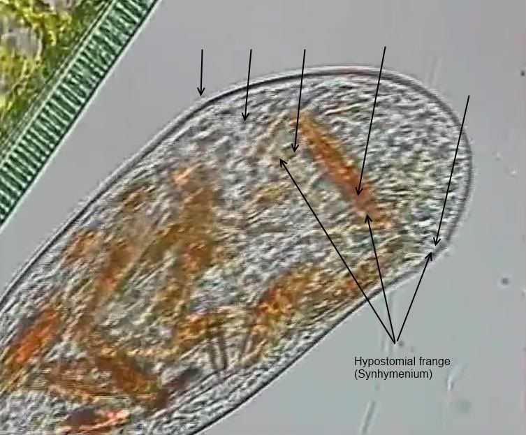
To resolve the identification below the order Synhymeniida you will need a clearer view of the surface ciliation...in particular, the synhymenium / hypostomial frange mentioned above, and any "sutures" where kineties meet.

These are beautiful images, and the cytopharyngeal basket is stunningly clear. :)

RogelioMoreno
Posts: 2979
Joined: Fri Nov 20, 2009 11:24 am
Location: Panama

Post by RogelioMoreno »

Bruce,

I will try to get images of the surface, I think there are more specimen in my sample.

Thank you very much for your comments and help.

Rogelio

RogelioMoreno
Posts: 2979
Joined: Fri Nov 20, 2009 11:24 am
Location: Panama

Post by RogelioMoreno »

Bruce,

I have not found another subject; but the following are pictures of a subject that I found before the posted above.

Image

Image

Image

Rogelio

Bruce Taylor
Posts: 827
Joined: Thu Jun 23, 2011 4:49 pm
Location: Wakefield, Quebec / Ottawa, Ontario
Contact:

Post by Bruce Taylor »

Hi Rogelio,

The second picture (from the new set) shows something that could be the synhymenium. However, the twisting and compression of the cell is confusing me...I can't tell what is where. If I were more experienced, I could probably unravel the puzzle.

What I was hoping to see was something like a line across the cell, in the vicinity of the oral aperture. Here's a still I took from one of Francisco's videos, which I believe to be Zosterodasys. The "frange" is very faint, but just visible...I've added arrows to identify it.

Image

The ends of the frange do not necessarily meet at the back of the cell, but may be offset. In Nassula, the frange is partial, running from the mouth to the right of the cell (admittedly, I have great difficulty even seeing it in brightfield, with all the colourful particles inside Nassula!).
Last edited by Bruce Taylor on Sun Apr 01, 2012 6:34 am, edited 2 times in total.

carlos.uruguay
Posts: 5358
Joined: Thu Feb 23, 2012 8:05 pm
Location: Uruguay - Montevideo - America del Sur
Contact:

Post by carlos.uruguay »

Maravilloso!
Realmente.
Wonderful. really

RogelioMoreno
Posts: 2979
Joined: Fri Nov 20, 2009 11:24 am
Location: Panama

Post by RogelioMoreno »

Bruce, thank you very much for the additional information.

Carlos, thank you.

Rogelio

Post Reply Previous topicNext topic