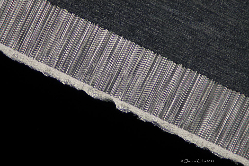
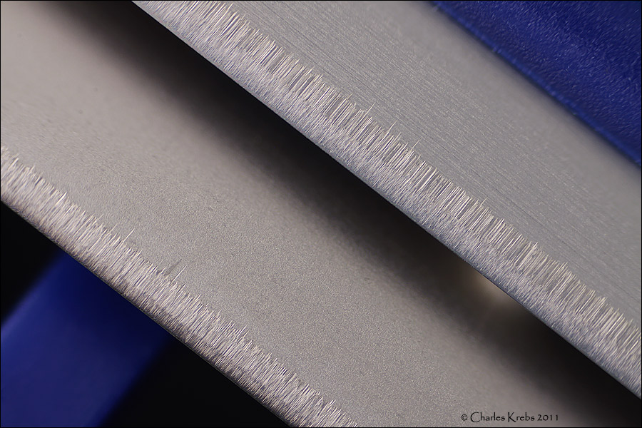
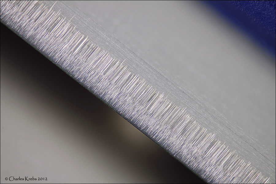
I'm using diamond paste on a glass plate and it's getting there but I have a long way to go.
Metallurgical specimens mounted into bakelite typically , when I did it early last century, were taken down through silicon carbide "wet and dry" paper to 400 grit, then diamond paste in usually only one grit size, which I think was 1 micron, (I forget but think the next, not often used - only for very hard specimens, was 0.1 micron.) That stage was pretty fast, held by hand on a disc covered in a cloth going round at a few revs per second, for a minute or so. The next stage was Alumina, much finer, something like 0.05micron. These finer stages were much faster than the "wet and dry" stages though, so I'm surprised that it's taking you a long time to polish your thin knife edge. SO what I'm getting round to - perhaps your grit size is wrong?
Our specimens were often quite chunky, such as sections of gas turbine blade or nuclear power station - or my motorcycle engine after it had been "tested" with nitro methane in the fuel, and blown to bits.
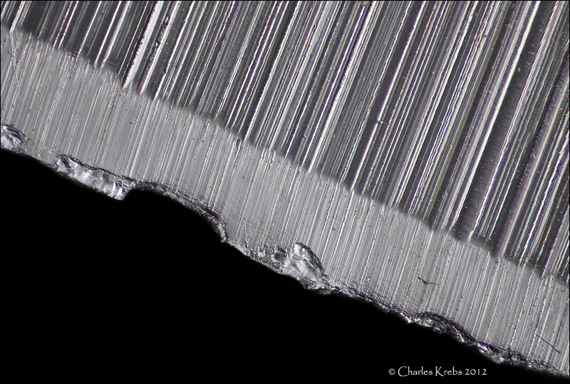
(The surface was ready for etching, to show the microstructure, as soon as it looked like a mirror. Remaining scratches were sometimes useful to indicate relative hardness of the phases in the specimen.
If you google "Steel microstructures" for the type (stainless, high carbon, etc) of the material you're using you'll probably find you need 40x and higher magnification, which is going to be tough. You would start to see how fine the structure is below that, without actually resolving it. An Iron carbon ( in steel) phase which has a lamellar structure was named "pearlite" early on. when microscopes couldn't resolve the detail. - it looked "pearly".
However if your knives have a surface treatment to help with the hardness, etching could show that up very well and it would be visible at more achievable magnifications. Case hardening, nitriding, titanium treatments etc could fall into this category.)