
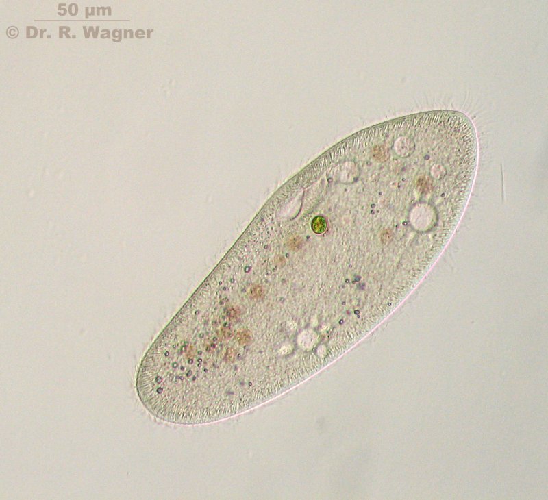
these are my first shots of a paramecium that really show some details.
You can see the 2 contractile vacuoles encircled by their feeding ducts, the oral groove with the formation of a new food vacuole at its end and the overall surrounding cilia.
The next phase-contrast picture lets us have a look at the macronucleus (red arrow). It is a dark oval partially hidden by a contractile vacuole. The yellow arrow in the inset points to what I am thinking of is the micronucleus. Does somebody know if that could be true?

In the first two pictures it was necessary to have the paramecium in a rather flat state under the coverslip (actually it died a few seconds after my last shot). Otherwise the shown details would have been unvisible. On the other hand it is interesting to show the spacial dimensions of that critter, too. This picture of a freely mobile and unflattened paramecium shows us that especially the oral zone is completely different now.


