Olympus BHS. Olympus 40/0.95 S Plan Apo. Olympus NFK 1.67x photoeyepiece. DIC illumination with electronic flash. Canon 50D.
Olympus BHS. Olympus 60/1.40 S Plan Apo. Olympus NFK 1.67x photoeyepiece. DIC illumination with electronic flash. Canon 50D.
Rotifer with egg amid Trachelomonas
Moderators: rjlittlefield, ChrisR, Chris S., Pau
- Charles Krebs
- Posts: 5865
- Joined: Tue Aug 01, 2006 8:02 pm
- Location: Issaquah, WA USA
- Contact:
- Ernst Hippe
- Posts: 205
- Joined: Sun Aug 16, 2009 9:41 am
- Location: Germany
-
- Posts: 2979
- Joined: Fri Nov 20, 2009 11:24 am
- Location: Panama
- Charles Krebs
- Posts: 5865
- Joined: Tue Aug 01, 2006 8:02 pm
- Location: Issaquah, WA USA
- Contact:
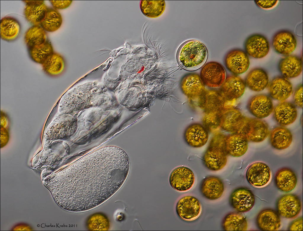
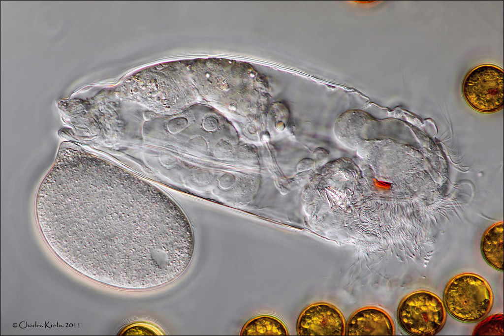
I'm not sure of the "composition" of rotifer eyes. More basic protozoan eye-spots are groups of lipid globules of carotenoid pigments. They provide some orientation to existing light and (as is clearly seen in trachelomonas and dinoflagellates) allow the organism to move toward the light.
In a discussion of whether or not to use eye-spots for classification purposes, there is some interesting information in this excerpt:
There are a couple of older shots here that show rotifer eye-spots clearly:
http://www.photomacrography1.net/forum/ ... php?t=4804
In a discussion of whether or not to use eye-spots for classification purposes, there is some interesting information in this excerpt:
There are a couple of older shots here that show rotifer eye-spots clearly:
http://www.photomacrography1.net/forum/ ... php?t=4804
- Wim van Egmond
- Posts: 826
- Joined: Tue Aug 15, 2006 9:28 am
- Location: Berkel en Rodenrijs, the Netherlands
- Contact: