Structured Illumination Microscopy (Zeiss Apotome)

A forum to ask questions, post setups, and generally discuss anything having to do with photomacrography and photomicroscopy.

Moderators: rjlittlefield, ChrisR, Chris S., Pau

Tardigrade37
Posts: 134
Joined: Tue Mar 04, 2008 7:38 pm

Structured Illumination Microscopy (Zeiss Apotome)

Post by Tardigrade37 »

I have posted some fluorescence images acquired with a structured illumination device known as the Apotome. I have alluded to how this works, but I would like to show you the process from start to finish. Essentially, a grid is placed in the illumination path and a set of three images is acquired when the grid shifts position. Then, using a proprietary algorithm, the images are reconstructed to remove out of focus light. All of this is controlled by the Zeiss software Axiovision. This generates a set of optical sections very similar to those acquired by a confocal laser scanning microscope. To learn more, click here.

Many have asked how long it takes to acquire an image, so I used a pollen slide to show you some examples. Here are the settings:
EXFO Excite 120W Metal Halide excitation
Rhodamine filter cube
Plan-Apochromat 63x/1.4 oil immersion objective
Hamamatsu ORCA-AG camera
Exposure time: 3ms
Averaging: 1
Number of images: 109
Z sampling: 275nm
Total acquisition time: approximately 8 minutes, so roughly 5 seconds for a single slice - this includes all of the overhead for opening/closing shutters, moving the focus, shifting the grid, and processing the three images.


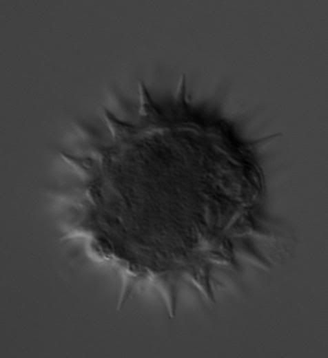
Here is a DIC image of the pollen:
Image


When the Apotome is engaged, you see this through the oculars:
Image


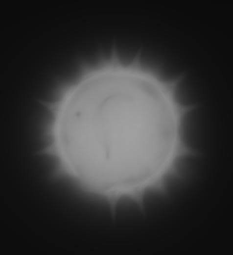
A conventional fluorescence image would look like this:
Image


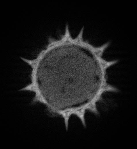
An optical section reconstructed from the three grid images looks like this:
Image


When the 109 images are volume rendered, you get something like this - not the best rendering, it was a quick and dirty job:
Image

Let me know if you have any questions!
Chris

lauriek
Posts: 2402
Joined: Sun Nov 25, 2007 6:57 am
Location: South East UK
Contact:

Post by lauriek »

Looks like a pretty cool setup you have!

I presume you could not use the Apotome with a conventional DSLR, is that right? (I'm assuming the Apotome sw has to control the camera...)

Tardigrade37
Posts: 134
Joined: Tue Mar 04, 2008 7:38 pm

Post by Tardigrade37 »

Hi Laurie,
I believe you can use any camera with this, so long as the drivers to control the camera are available. I know there is a module for Axiovision that will control Canon cameras, but I've never used it.

That being said, I can't see much of an advantage to using a DSLR. For one, the bayer filter in front of the CCD would decrease spatial resolution as the fluorescence through these filters is generally monochromatic. The Hamamatsu camera I am using is cooled to -30 °C, thus greatly reducing spurious thermal noise. It also has a much higher quantum efficiency for capturing precious fluorescence photons. Lastly, the Apotome uses two different grids for the high and low magnification ranges. I only have the high mag. grid, so I can only use the 63x and 100x objectives with it. According to the table below, the 1344x1024 resolution of the Hamamatsu is suitable for capturing all of the detail in an image using a 1.0x coupler.

Image

Post Reply Previous topicNext topic