Hi Charlie
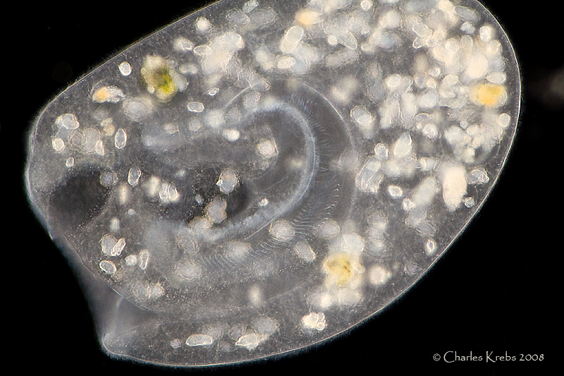
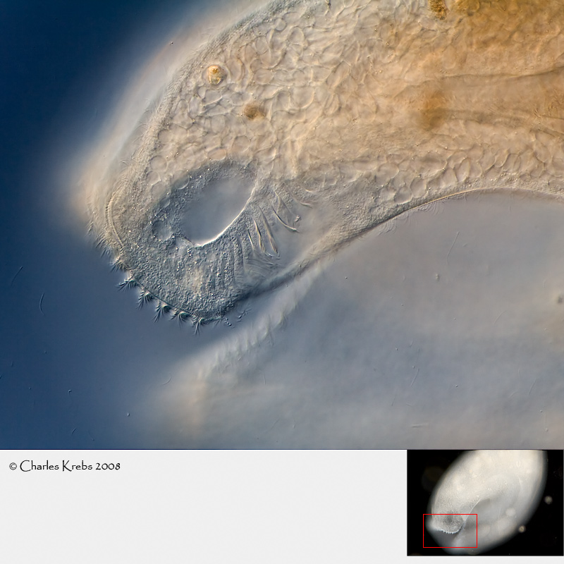
I would like to use your post with your excellent images of Bursaria truncatella as an opportunity to show some images of my own collection with the intention to show some further details of the cell surface of this giant species.
This is the cell surface of Bursaria truncatella taken with oil immersion (believe me). Try to imagine the image without the yellow labelling. At first glance it seems to be a little chaotic without any hints of further charateristics of Bursaria. But the areas in boxes offer some new features.

In the first box three contractile vacuoles (KV) in various stages of activity are visible. The complete body of B. truncatella is covered with hundrets of small contractile vacuoles. In a free swimming cell they cannot be recognized. Top right a diastole. Top left a systole and EP is the excretion pore of a contractile vacuole.

The box 2 shows that the ciliary rows (kineties) of B. truncatella consist not of singular cilia but of pairs of them. Usually this feature is only visible after silver impregnation. At this magnification I could just separate them (arrows).

In box 3 the mitochondria (MI) beneath the pellicle of B. truncatella are visible. They provide the energy for the movement of the cilia.
http://www.einzell.de/Forum/Bursaria-4-P4132953.jpg
I hope you agree that I used your post as an vehicle for this images!
Martin