I collected some bright green "slime" and water from a very slow moving creek, hoping to get some subjects for the microscope. The "green" was thousands of Closterium, a crescent-shaped desmid (a single-celled green algae). The next morning I was surprised to find numerous hydra in the sample as well. Some were suspended from the water's surface. So I put a few in a small "tank" and took a couple macro shots. It was interesting to watch the method they used to get to the surface. They would latch onto a bubble or two and use them to float up to the surface, where they would then spread out looking for a meal in this upside-down position.
Reversed 50/2.8 El Nikkor on bellows, Nikon D200.
Minolta 12.5/2 Macro on bellows, Nikon D200.
Hydra and Closterium
Moderators: rjlittlefield, ChrisR, Chris S., Pau
- Charles Krebs
- Posts: 5865
- Joined: Tue Aug 01, 2006 8:02 pm
- Location: Issaquah, WA USA
- Contact:
- rjlittlefield
- Site Admin
- Posts: 23625
- Joined: Tue Aug 01, 2006 8:34 am
- Location: Richland, Washington State, USA
- Contact:
- Planapo
- Posts: 1583
- Joined: Tue Nov 07, 2006 2:33 am
- Location: Germany, in the United States of Europe
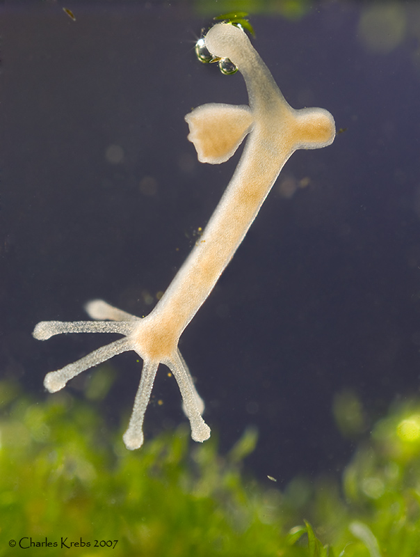
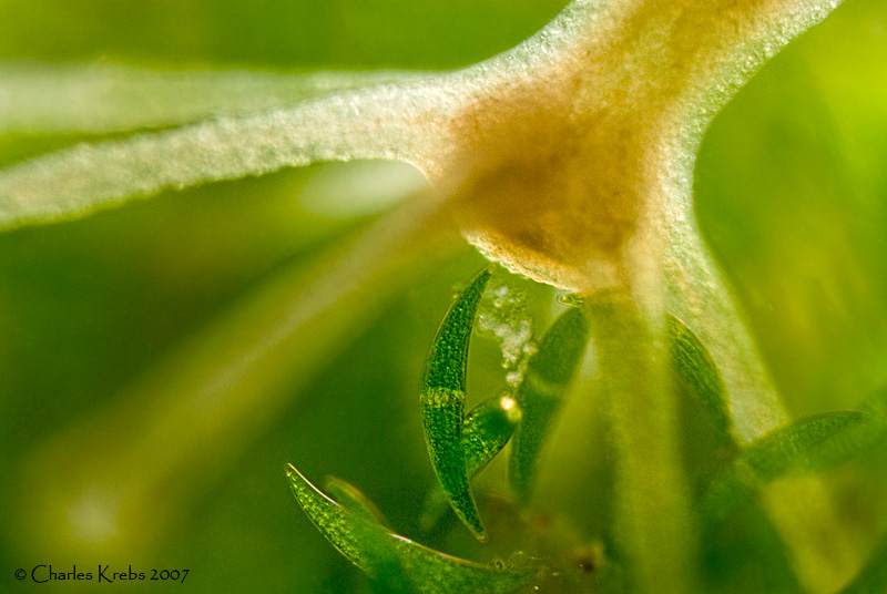
Yes, very beautiful, Charlie!
You manage to shoot highly informative and at the same time aesthetically appealing photos even in such situations that I would find too tricky to tackle.
Did you have your camera mounted vertically on a repro stand or tripod for this and was it lit by flash?
Looking forward to enjoying more of your inspiring photomacrography.
--Betty
I remember from biology class that hydras reproduce with budding. I like the first picture because you can see it at the base of the bigger one. OK maybe you see that thousands of times.
And the second with the algae is also very interesting, and most unusual. Do you know the scale of this picture?
Péter
- Charles Krebs
- Posts: 5865
- Joined: Tue Aug 01, 2006 8:02 pm
- Location: Issaquah, WA USA
- Contact:
Rik... No stacking possible here. Although they don't move very fast they were constantly moving. The upper image was from two, manually combined (roughly top half and bottom half). The lower is a single image.
Betty... this is my "tabletop" setup, still pretty much as pictured in the technique. (Horizontal bellows on top of Olympus microscope focus block). I did use electronic flash.
Péter... I chose this particular specimen to photograph specifically because of the "budding" that can be seen. The Closterium, seen more clearly in the lower picture, average about .6mm (.024 inch) in the long dimension (measured along a straight line between the two tips). They average about .08mm (.0032 inch) diameter at the thickest section at the middle.
I hope to get around to putting a couple microscope Closterium shots over in the microscope section. If/when I do I'll mention it here.
- Charles Krebs
- Posts: 5865
- Joined: Tue Aug 01, 2006 8:02 pm
- Location: Issaquah, WA USA
- Contact:
Laurie... yes, this is a relatively "huge" subject!
If you look closely in the "blue" area of the top shot you can see a diatom in the upper left (dark) and one in the lower right (bright, smaller). These diatoms are more the size subjects that get placed under the microscope (and these are pretty large as far as diatoms are concerned).