Salt water amphipod detail. Cross polarized illumination.

Images made through a microscope. All subject types.

Moderators: rjlittlefield, ChrisR, Chris S., Pau

Charles Krebs
Posts: 5865
Joined: Tue Aug 01, 2006 8:02 pm
Location: Issaquah, WA USA
Contact:

Salt water amphipod detail. Cross polarized illumination.

Post by Charles Krebs »

A while ago I was trying a variety of little critters with cross polarized light to view muscle structure. I had collected both saltwater and freshwater amphipods. Curiously the carapace of many of the saltwater specimens displayed a very interesting pattern in this light. This was not visible in non-polarized light. I looked for similar patterns on the freshwater species but did not find it on any.

Anyway, I thought it made an interesting abstract pattern...


Image

bernhardinho
Posts: 563
Joined: Sun Aug 13, 2006 6:28 am
Location: Germany
Contact:

Post by bernhardinho »

Hi Charles

no wonder you don`t see these crosses in non polarized light, because they show the planes of vibration of the polarizers themselves!! Just like in conoscopic observation:

http://micro.magnet.fsu.edu/primer/tech ... intro.html

When turning the polarizers while observing you can see these extinction crosses moving apart!

Bernhard

rjlittlefield
Site Admin
Posts: 23608
Joined: Tue Aug 01, 2006 8:34 am
Location: Richland, Washington State, USA
Contact:

Post by rjlittlefield »

Bernhard,

I am a little confused, and I wonder if you can help me understand.

This is the first time that I have encountered this term, "conoscopic observation".

At the link you provide, Figure 2 shows "Conoscopic Interference Patterns" that look just like the ones in Charlie's amphipod.

But the description there says that "Figure 2 illustrates conoscopic images of uniaxial crystals observed at the objective rear focal plane", as through a Bertrand lens. This is consistent with other articles I found about conoscopic inspection.

Charlie's picture, on the other hand, seems to have been taken with conventional optics (no Bertrand lens) and the cross pattern is observed in the image plane of the subject.

My mind is full of questions...

- Have I misunderstood the descriptions of conoscopic inspection?
- Is the conoscopic cross pattern also commonly observed in the image plane of the subject, and at multiple places at once? If so, then why do the descriptions emphasize use of a Bertrand lens?
- Most areas of the carapace do not show the cross pattern, and the areas that do, appear to have hard boundaries. Physically/chemically, what is different about the areas that show the cross pattern? Why does it appear only in the salt-water specimens?

Any further explanations or links that you can provide would be greatly appreciated.

Thanks!
--Rik

bernhardinho
Posts: 563
Joined: Sun Aug 13, 2006 6:28 am
Location: Germany
Contact:

Post by bernhardinho »

Hi Rik

very good question. Did you read the full article in the link? Right towards the bottom of the page you find the polymers with part of the answer to your question.
I was trying to say that the cross is identical with the vibration planes of the polarizers JUST LIKE in conoscopical view. Of course Charles pics are in orthoscopical observation and the reason WHY this so-called spherulites show is different in biological organic objects compared to uniaxial crystalls! ( In fact spherulites belong to the world of polymers, but the term is used in botanics in analogy, at least as spherulitic structures!)
In the former the cross ( also referred to as Brewster's cross) indicates spherical anisodiametric structure of the fibrills that form the object. Very familiar examples are starch grains, tracheids or bordered pits in wood anatomy.

I don't think, by the way, that they show in salt water specimens only. This may be pure chance.

Oh Lord, that is hard stuff to explain in English!!!


Bernhard

rjlittlefield
Site Admin
Posts: 23608
Joined: Tue Aug 01, 2006 8:34 am
Location: Richland, Washington State, USA
Contact:

Post by rjlittlefield »

Oh Lord, that is hard stuff to explain in English!!!
No doubt, but I appreciate the effort and I think you were very successful. :D

No, I had not read the article completely -- just quickly focused on the sections about "conoscopic". Thanks for the pointer to the part about polymers near the bottom of page.

I think I can puzzle it out from here.

Many thanks!

--Rik

bernhardinho
Posts: 563
Joined: Sun Aug 13, 2006 6:28 am
Location: Germany
Contact:

Post by bernhardinho »

Oh, by the why

I overlooked one of your questions and that is WHY the patterns on the carapace show at all.

Well I sort of ignored the question on purpose :oops: 'cause I don't know anything about amphipods. I didn't even know the word!

So hopefully someone comes up with an explanation for that one!


Bernhard

Charles Krebs
Posts: 5865
Joined: Tue Aug 01, 2006 8:02 pm
Location: Issaquah, WA USA
Contact:

Post by Charles Krebs »

The "Maltese cross" pattern is very reminiscent of those obtained viewing starch grains. I have no idea if this is found on one particular species or if it is a more common trait of saltwater (as opposed to freshwater) species. In any event the polarized light showed some structure that was not observed readily in "regular" lighting. Another "difference" I found when working with these samples was a very different appearance to the eyes between salt/fresh samples. I really know nothing about these species, so these are just very superficial observations. But the eyes were quite interesting in and of themselves, and perhaps I should collect some samples and see what I can do. (Problem is this would best be done with fairly high mag "epi" illumination, which I don't have)

gpmatthews
Posts: 1040
Joined: Thu Aug 03, 2006 10:54 am
Location: Horsham, W. Sussex, UK
Contact:

Post by gpmatthews »

I have played around with crystals of waxy films, both professionally and for fun. If you look at my pages at http://www.gpmatthews.nildram.co.uk/mic ... xtals.html
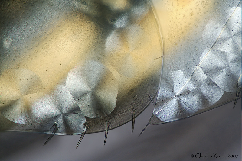
you can see at the bottom of the page some pictures of PEG3350 and a film of Gelucire 44/14, an amphiphilic wax. Both these show similar rosette structures in polarised light and the Gelucire shows maltese cross markings. Similar structures can also be seen in films of fatty alcohols and many other waxes. This appears due to the fact that waxes crystallise in spherules, which in films are constrained to be flat discs. Starches show a similar radial structure. I wonder whether the structures seen in the amphipod are due to a layer of waxy material, or whether they are characteristic of the chitinous exoskeleton. If the latter, it may be that deposition of chitin is different in salt water, if the former, it may be a specific adaptation to the salty environment. It would be interesting to compare other creatures...

The meeting of the rosettes show flattened boundaries. This suggests that the material grew on the chitinous substrate until it met its neighbouring rosette. Hence the substrate would exist before the observed structures were formed. This would support the wax-film hypothesis.

All speculation, I'm afraid, but you may find it interesting to prepare films of waxes by melting a speck between a slide and coverslip, then cooling (rate of cooling can be important) and examining in polarised light.
Graham

Though we lean upon the same balustrade, the colours of the mountain are different.

rjlittlefield
Site Admin
Posts: 23608
Joined: Tue Aug 01, 2006 8:34 am
Location: Richland, Washington State, USA
Contact:

Post by rjlittlefield »

Might one get some more info by washing the (poor dead) creatures in a solvent that dissolves wax, and seeing if the pattern goes away or at least diminishes?

--Rik

Charles Krebs
Posts: 5865
Joined: Tue Aug 01, 2006 8:02 pm
Location: Issaquah, WA USA
Contact:

Post by Charles Krebs »

On one hand it's a little embarrassing not knowing more about some of my subjects. But then I realize that this is one of the aspects of photomicrography that makes it so enjoyable... seeing things I (personally) have not seen before and then trying to learn more about them.

Here's a shot I had that shows the eye structure I thought would also be interesting to look at in more detail.

Image

Post Reply Previous topicNext topic