Daphnia longispina

Images made through a microscope. All subject types.

Moderators: rjlittlefield, ChrisR, Chris S., Pau

bernhardinho
Posts: 563
Joined: Sun Aug 13, 2006 6:28 am
Location: Germany
Contact:

Daphnia longispina

Post by bernhardinho »

Hi everbody,

are you still out there? It's been a bit quiet lately.

Here is Daphnia longispina

head and abdominal region (with claw). The last pic is a stack of three pics with Helicon focus.

Hope you enjoy:

Image

Image

Image


So long

Bernhard

Bruce Williams
Posts: 1120
Joined: Mon Oct 30, 2006 1:41 pm
Location: Northamptonshire, England
Contact:

Post by Bruce Williams »

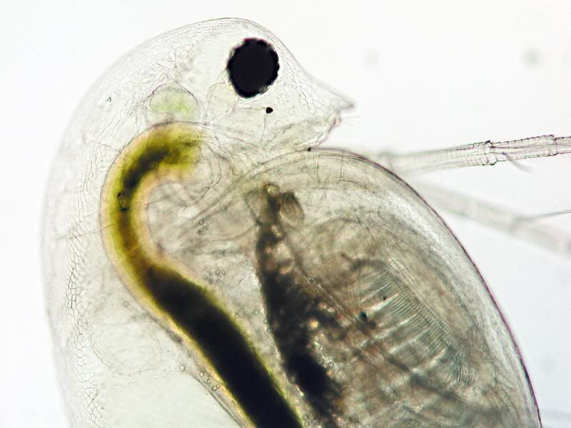
I just had to reply to this posting Bernhard - VERY impressive! Three excellent images - all showing an astounding amount of internal detail - worth taking time to really explore and enjoy.

Thanks.

Bruce :D

bernhardinho
Posts: 563
Joined: Sun Aug 13, 2006 6:28 am
Location: Germany
Contact:

Post by bernhardinho »

Thanks, Bruce,

if you're prepared for even more, take a look here:

http://www.mikroskopie-forum.de/read.ph ... #msg-29164


Bernhard

rjlittlefield
Site Admin
Posts: 23622
Joined: Tue Aug 01, 2006 8:34 am
Location: Richland, Washington State, USA
Contact:

Post by rjlittlefield »

Bernhard, these are beautiful pictures!

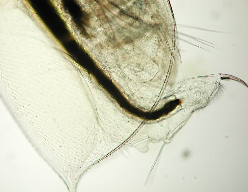
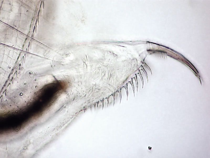
I am interested in the texture of the creature's "shell". Somehow I expected it to be smooth and featureless, but it appears to be a fairly regular pattern of tiled segments. Do you know how that structure forms? Is it for example one tile per cell, or is the structure much larger or much smaller than the level of a cell?

--Rik

bernhardinho
Posts: 563
Joined: Sun Aug 13, 2006 6:28 am
Location: Germany
Contact:

Post by bernhardinho »

Very good question, Rik

first of all, you will hardly find smooth structureless shells in crustacea. In fact, these patterns are an important feature for the ID.
I found the answer to your question in one of our standard books over here (Das Leben im Wassertropfen- Life in a waterdrop):

For those who understand german:

" Die feinen, polygonalen Muster der durchsichtigen Schalenkutikula spiegeln die Grenzen der unter der Kutikula liegenden Hypodermiszellen"

For those who prefer english:

" The fine polygonal patterns of the transparent shell cuticula are indicating the outlines of the hypodermic cells beneath the cuticula."


I hope that makes things clearer!

Bye

Bernhard

Wolfgang Bettighofer
Posts: 58
Joined: Sun Mar 11, 2007 1:42 pm
Location: Kiel, Germany

Post by Wolfgang Bettighofer »

Hi Bernhard,

high quality pictures combined with an interesting discussion about details of crustaceae anatomy.

Thank you!

--Wolfgang

rjlittlefield
Site Admin
Posts: 23622
Joined: Tue Aug 01, 2006 8:34 am
Location: Richland, Washington State, USA
Contact:

Post by rjlittlefield »

bernhardinho wrote:I hope that makes things clearer!
Yes, wonderfully so! Thanks! :D

--Rik

Bruce Williams
Posts: 1120
Joined: Mon Oct 30, 2006 1:41 pm
Location: Northamptonshire, England
Contact:

Post by Bruce Williams »

Bernhard - Thanks for the link to your other D. longispina pics on the German forum . More excellent photos - I strongly recommend anyone to pay a visit.

Bruce

beetleman
Posts: 3578
Joined: Fri Aug 04, 2006 4:19 am
Location: Southern New Hampshire USA

Post by beetleman »

They look like x-rays of an alien creature. Wonderful pictures and Info.
Take Nothing but Pictures--Leave Nothing but Footprints.
Doug Breda

Post Reply Previous topicNext topic