
In that post, I showed some feeding behavior that I had not seen before, in which the bug removed some of its mouthparts from the rostrum while probing for food. At the time, I thought those mouthparts were the feeding tube. But on further investigation, I learned otherwise.
Starting at the end, here is a detail of the tip of the feeding tube.
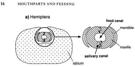
Quoting here from R.F.Chapman, "The Insects / Structure and Function" (pages 15-16),
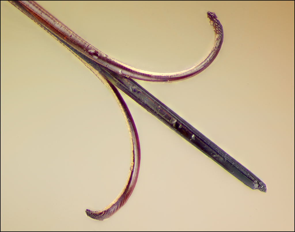
Image #1 at top of post shows a prepared specimen, in which the maxillary and mandibular stylets were pulled away from the labium and separated slightly as they dried. The barbed ends of the mandibular stylets can be seen clearly as the curled structures, while the interlocked maxillae remain straight at the center of the assembly.In Hemiptera, mandibles, maxillae and labium are all elongate structures, while the labrum is relatively short. The food canal is formed by the opposed maxillae which are held together by a system of tongues and grooves. These allow the stylets to slide freely on each other, while maintaining the integrity of the food canal. The maxillae also contain the salivary canal. On either side of the maxillae are the mandibular stylets. These are the principal piercing structures and they are often barbed at the tip. When the insect is not feeding, the slender maxillary and mandibular stylets are held within a groove down the anterior side of the labium. The hemipteran labium is known as the rostrum. It is usually segmented, allowing it to fold as the stylets penetrate the host.
Here are two more images showing a wider view for context.
Image #2 is the prepared specimen again. The labium (rigid part of the rostrum) is held out of the way with an insect pin, while the labrum and stylets are in more or less their normal position except for the curling of the mandibular stylets.
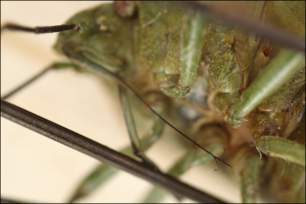
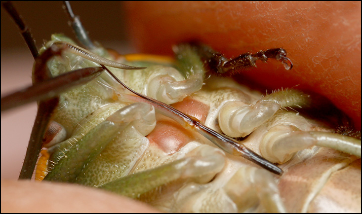
Image #3 is a different specimen, this one live, using an insect pin to manipulate the mouthparts so as to show the labrum and the stylets in relation to the rigid segmented labium that forms the rostrum.
You can see another view of these structures in operation in THIS POST by Mike Gordon at flickr.
I hope you find this interesting!
--Rik
Technical details...
Image #1: Nikon M Plan 20X NA 0.40 ELWD objective, 55 frames in Zerene Stacker, PMax + DMap combined. Illuminated by Canon 580EX II Speedlite plus two Yongnuo 460 II flashes through paper diffusers.
Image #2: Olympus 38 mm f/2.8 bellows macro lens at f/5.6, same illumination.
Image #3: Canon MP-E 65 at ~2X, f/11, single 580EX II Speedlite with Opteka diffuser.
