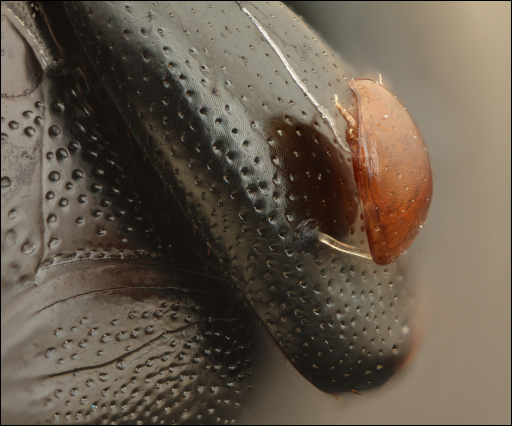
This tiny brown bug is firmly attached to the leg of a small black burying beetle. I didn't know the beast was there when I gave the black beetle a good wash in an ultrasonic cleaner. After the bath it's still there. I think the white fiber on the beetle's leg behind it is not related, but I'm not sure.
Frame height ~1.5 mm.
Can somebody please tell me what this is?
--Rik
Setup: Mitutoyo M Plan Apo 10X NA 0.28 with MT-1 tube lens on Canon T1i, single flash diffused through foam cup, Zerene Stacker DMap+PMax, 81 frames at 0.010 mm focus step on StackShot.
Edit: change title to reflect ID
Last edited by rjlittlefield on Thu Apr 26, 2012 1:43 pm, edited 1 time in total.
No ID here Rik, but you make me curious on your washing method.
Never heard of someone using an ultrasonic bath on bugs before.
In which (combination of) fluid(s)? I can imagine it's not suitable of every kind of species.
This was done with a general purpose solution sold specifically for use diluted with water in an ultrasonic cleaner. It works well for simple debris and light degreasing, for subjects that play well with water.
Hairy subjects are likely to clump. Some of those will come back OK when dry by fluffing with an air jet or gentle brushing, but some such as the barbed hairs of many bees will stick together stubbornly. (I was recently told by an entomologist friend that such critters may fluff out again when bathed in chloroform and re-dried, but I haven't tried that.)
I have also been told that rarely a specimen will essentially explode when the ultrasound is turned on. I presume this is because the specimen is particularly fragile and dry, so it just gets shaken apart. Some quiet soaking beforehand seems like a good idea to both prevent breakage and soften any debris to be removed.
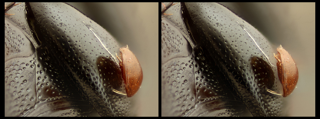
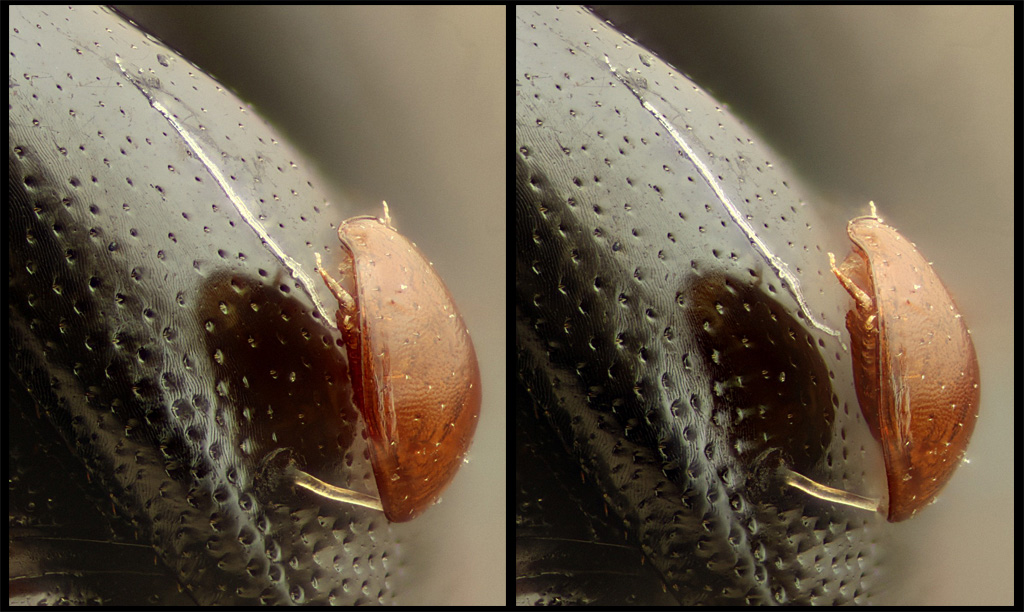
Here are a couple of stereo renderings that may help to appreciate the structure.
I switched objectives and changed the viewpoint slightly because I was exploring some other issues. These were shot with a Nikon CFI 10X NA 0.25, processed with PMax hence the higher contrast.
So then a good reference for more info is perhaps this article: http://hiltonpond.org/ThisWeek040508.html, which discusses a mutually beneficial relationship between the mites and the beetles.
The mite measures 0.65 mm end to end as seen in the photo. The upper frame of the stereo pairs is full frame on a Canon T1i, 22.3x14.9 mm sensor, at 10X.
rjlittlefield wrote:The mite measures 0.65 mm end to end as seen in the photo. The upper frame of the stereo pairs is full frame on a Canon T1i, 22.3x14.9 mm sensor, at 10X.
It is a mite. The legs look like those of Astigmata but the high degree of sclerotisation suggests other possibilities. That pale tube is unsegmented and unlikely to be part of the mite, unless it is everted gut. It might be a parasite but that seems unlikely. I await the specialist comment with interest.
Harold
My images are a medium for sharing some of my experiences: they are not me.
This is an email response I got from my friend, Josh Vlach, our mite specialist here at Oregon Department of Agriculture.
"I wouldn't call myself a mite specialist, but here is my answer anyway. I don't believe this is the 'typical' phoretic mite associated with carrion beetles. Although I won't try and identify this mite from a picture, this appears to be a type of mite that uses insects (usually beetles) as simple transport and does not have the more developed symbiotic relationship with the beetle. I don't believe the pale, waxy appearing line is related to the mite (but there are plenty of behaviors that are yet unobserved). The strand that attaches the mite to the beetle is exuded from the anus and is very durable and flexible. The mite is traveling in an immature stage (usually deutonymph). When it arrives at its destination (who knows how IT knows), it dissolves the strand. I've often thought someone should research its composition to make a better glue. Since I can't tell what type of mite it is, it is worth mentioning that there are other mites that have suction cups around their anus to attach to an insect. They often don't have functional mouthparts or digestive tract. Oddly, they feed on their insect host through their anus.
Enjoy,
Josh"
Steve
"You can't build a time machine without weird optics"
Steve Valley - Albany, Oregon