 Metal-Halogen Biomaterials by mygale
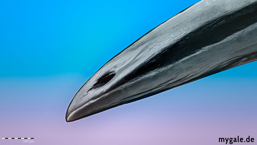
Ventrally on the chelicerae are the fangs, these hollow hypodermic needle-like structures are what delivers the venom into prey or attackers. Shown here is the tip of a tarantula fang, including the single opening of the venom canal on the dorsal side around 700µm from the tip away.
As with hypodermic needles, scorpions and venomous snakes the venom canal never ends at the tip itself, but shortly before it. This prevents the venom canal from getting clogged and is mechanically more stable. Those multi-use injection needles must be sufficiently tough to withstand the initial impact of a rapid attack, while at the same time they need to be hard and stiff to be able to break the prey’s protective cuticle. Shortly after the molt, they are white and vulnerable, tarantulas can’t use them to eat or perform other tasks, it takes a couple days to harden the fangs. A juvenile Chromatopelma cyaneopubescens (Strand, 1907) after the molt: https://flic.kr/p/D5CEXT
So what makes them strong enough the brake insect cuticle, which is essentially the same material? The spider takes advantage of a wide range of the available chemical and structural modifications in its cheliceral fangs. One of the secrets are the metals. The tarsal claws, chelicerae, stings and other tools of arthropods contain extraordinary amounts of heavy metals (e.g., zinc, manganese, copper) and halogens like bromine and chlorine. Those Metal–halogen biomaterials are widely distributed, especially among arthropods. The functions of the enriched structures suggest that metal–halogen biomaterials enhance mechanical properties. It is likely that they affect the behavior and ecology of the large fraction of arthropods in which they are found (Schofield, 2005). For example, leaf cutter ants may delay leaf cutting until zinc has hardened their mandibles (Schofield et al. 2002). The same effect may apply to tarantula fangs, as shown above. Other secrets of the strength are the chitin and its structure, the proteins and the water content (Politi et. al. 2012).
Technical info: 0.1 Acanthoscurria geniculata fang (chelicerae) - Sony Alpha 7RII + Cognisys StackShot + Nikon 10x MRL00102 @ ~10x Stacked from 156 images (8 µm steps), Helicon A + B. Ikea Jansjö, foam cup DIY diffuser + reflector, 9500px*5300. 50 Megapixel.
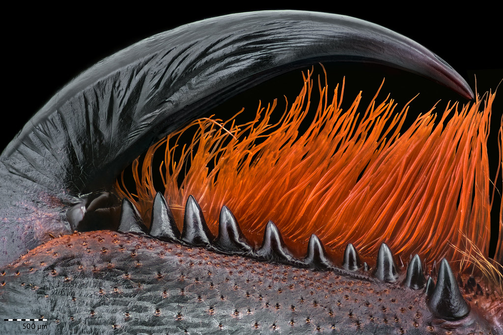 Tarantula fang (Chelicere) @6,2x by mygale
Situated prolaterally on the chelicerae are the cheliceral teeth. The cheliceral teeth aid in helping the spider crush its food. Strikers situated retrolaterally on the chelicerae (Image: https://flic.kr/p/U9zSaA ) form half of the primary stridulating organ, which is rubbed against paddle setae on the maxillae to produce sound. Check the Anatomy Poster for the rest of the text.
Technical info: Psalmopeus irminia Saager, 1994: cheliceral teeth / fang Sony Alpha 7RII + Cognisys StackShot + Nikon 10x MRL00102 @ ~6,2x Stacked from 145 images (20µm), Helicon B + CCC. 3x Ikea Jansjö, foam cup DIY diffuser + reflector, 8115px*5500.
 Theraphosa blondi palpal bulb @6x (test) by mygale
In mature males, the pedipalps host the male's copulatory organs - the palpal bulb. At the base you have the subtegulum, connected to this is the tegulum, which comprises the lower half of the palpal bulb. The bulb then thins into the emboli (shown in this image). This is the part of the bulb, which injects the sperm into the female‘s spermathecae. Many species have keels on their bulbs and these can present in a superior or inferior fashion. Check the Anatomy Poster for more information.
Testing the 10x Nikon at 6x with the Raynox 250 (normal position) as tube lens. Sony Alpha 7RII + Cognisys StackShot + Nikon 10x MRL00102 @ ~6,2x Stacked from 53 images (30µm; 50µm and 40µm didn’t work so well), Helicon B + C. 3x Ikea Jansjö, bendable concave DIY diffuser + reflector, 40 Megapixel. v1.1
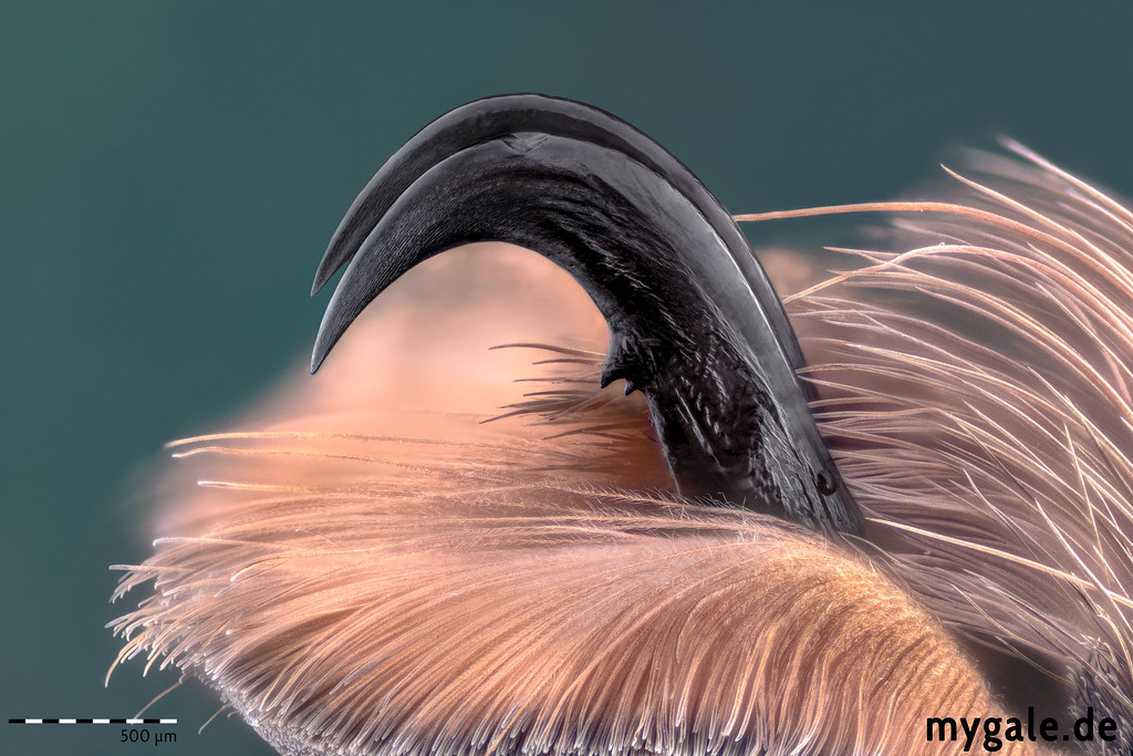 Tarantula Tarsal Claws 10x by mygale
Claws at the distal end of the tarsi. Female spiders of some families possess 1 single claw on the pedipalps, but these are no theraphosids. Some theraphosid genera, however, show a poorly developed third claw. All web-building spiders have three claws, as it is used to grab the silk strand, but not all three-clawed spiders build webs. Check the Anatomy Poster for more information.
P. subfusca Tarsal Claws LIII Deep Stack @ ~10x. Stacked from 300 images (9µm), Helicon B (18/4) + B (40/4) + C + C Slab (14). Flash Setup, 42 Megapixel. 35 GB Raw files + 100 GB tif files. About 6h post processing. v1.0
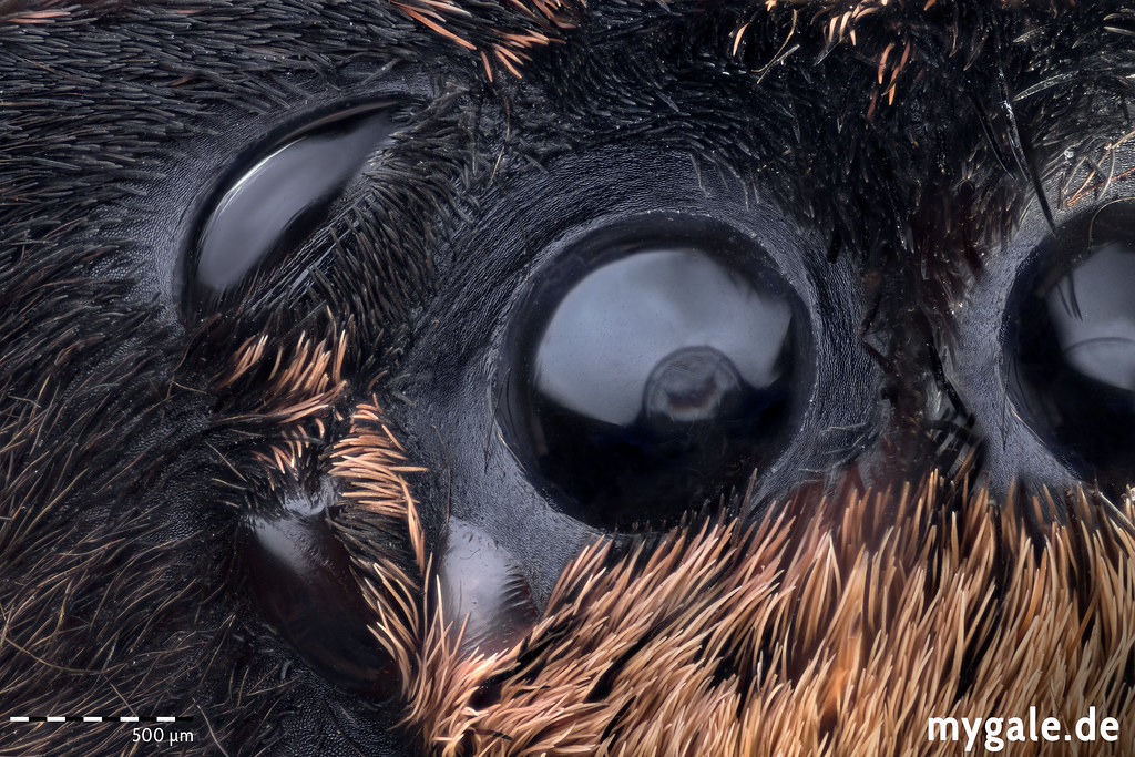 Tarantula eyes 10x by mygale
Theraphosids possess eight eyes arranged into two rows evenly. The top row features the anterior eyes whereas the bottom hosts the posterior. The middle four eyes, that is two on each row are the anterior and posterior medial eyes, respectively. Whereas the outer four eyes, also two of each row are the anterior and posterior lateral eyes. The secondary eyes have a tapetum lucidum - which reflects light. Therefore the primary eyes are used in conditions of better lighting whereas the secondary eyes can aid in vision in darker conditions. Theraphosids have very basic ocelli but they can see in wavelengths of 550 - 640nm based on a study of their photoreceptors by Dahl & Granda (1989)… Check the Anatomy Poster for more information.
Subject: P. subfusca ocular tubercle Deep Stack @ ~10x. Stacked from 170 images (8µm), Helicon B (8/4)+ B (16/4)+ B (40/4)+ C. Flash Setup, 35 Megapixel. The third test with the Nikon.
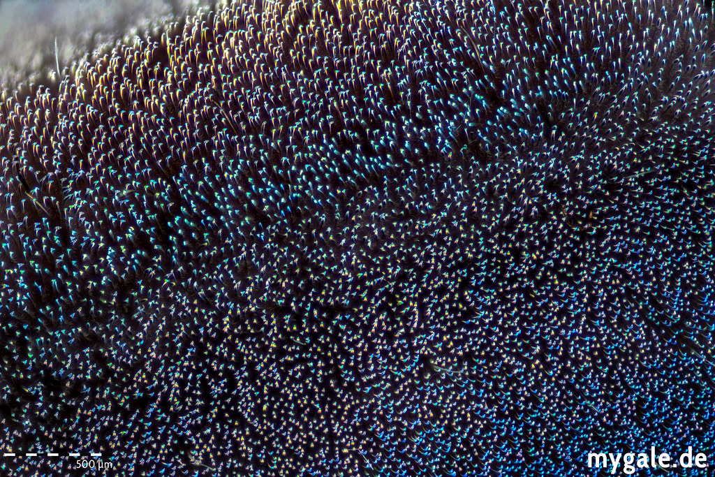 Scopulae @ 10x by mygale
Subject: (P. irminia L2 metatarsus ventral) On the tarsi and metatarsi of conventional theraphosid specimens are the scopulae located - specialized adhesive pads - which consist of very fine microscopic hairs, which enable the spiders to efficiently climb surfaces. The presence/absence of metatarsal scopulae and the extent of scopulation are two stable characteristics in theraphosid taxonomy.
Sony Alpha 7RII + Cognisys StackShot + Nikon 10x MRL00102 @ ~10x Stacked from 106 images (9µm), Helicon C only. Flash Setup, 40 Megapixel.
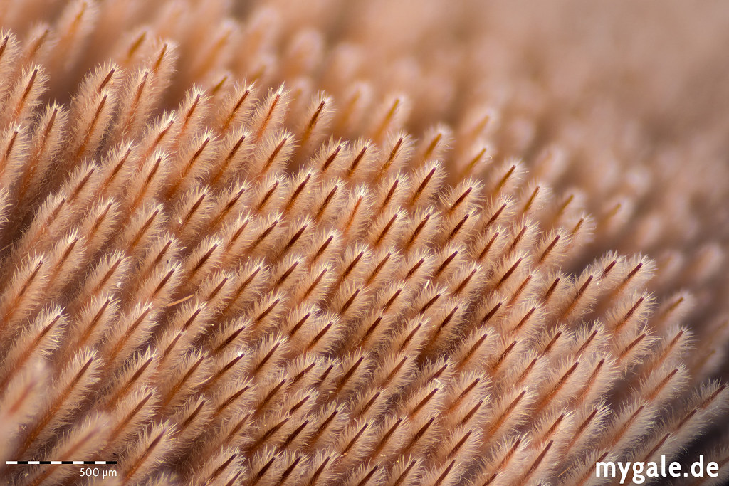 Plumose specialized setae @ 10x by mygale
Subject: Plumose specialized setae on the retrolateral side of the chelicerae from C. marshalli. Strikers situated retrolaterally on the chelicerae form half of the primary stridulating organ, which is rubbed against paddle setae on the maxillae to produce sound. In some species the strikers are absent and instead scopulae, made of plumose specialised setae is found on the chelicerae and thorn like structures on the maxillae and this produces sound just like the more conventional stridulation organ. Not all genera that stridulate have these forms of stridulation. Check the Anatomy Poster for more information.
Sony Alpha 7RII + Cognisys StackShot + Nikon 10x MRL00102 @ ~10x Stacked from 106 images (9µm), Helicon C only. Flash Setup, 39 Megapixel.
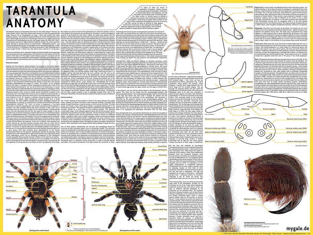 Tarantula Anatomy Poster (v1.0) by mygale