Hello,
here is part II of Plagiomnium ellipticum:
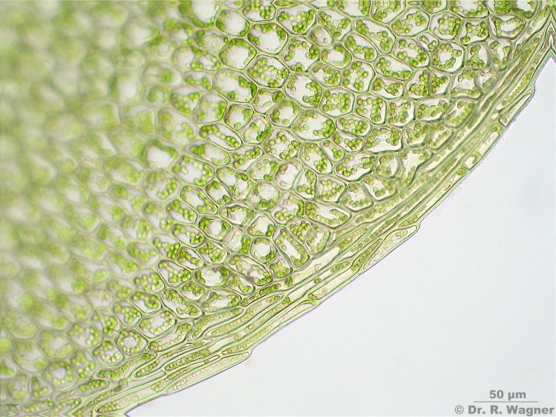
d) Seamed leaf border:
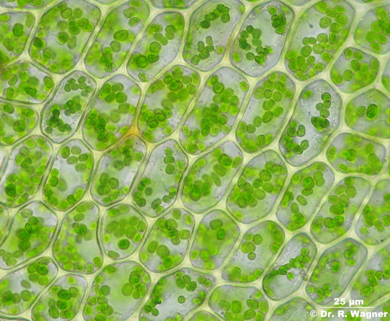
e) Polygonal lamina-cells. On a closer look at the chloroplasts you will even find some of them in dividing-process:
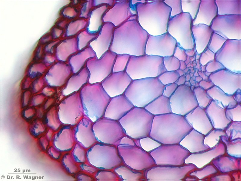
f) Stem, cross section in Etzold stain. Central part with central string (hadrom) and surrounding conducting tissue. At the border we see the outer cortex-layer (stereom). This is the way the stem of most leaf-mosses (Bryopsida) looks alike:
Enjoy!
Plagiomnium ellipticum, Part II
Moderators: rjlittlefield, ChrisR, Chris S., Pau
- ralfwagner
- Posts: 441
- Joined: Sat Aug 05, 2006 8:34 am
- Location: Germany, Duesseldorf
- Contact:
Plagiomnium ellipticum, Part II
----------------------
Ralf
http://www.lichenes.de
http://www.dr-ralf-wagner.de
http://www.youtube.com/user/drralfwagner
Ralf
http://www.lichenes.de
http://www.dr-ralf-wagner.de
http://www.youtube.com/user/drralfwagner
- rjlittlefield
- Site Admin
- Posts: 23606
- Joined: Tue Aug 01, 2006 8:34 am
- Location: Richland, Washington State, USA
- Contact:
- ralfwagner
- Posts: 441
- Joined: Sat Aug 05, 2006 8:34 am
- Location: Germany, Duesseldorf
- Contact:
Hello Rik,
look at pic a): There you see the leaf attached to the stem. Pic f) shows a cross-section of that stem. In order to obtain more contrast, this cross-section was stained with a mixture of some special blue and red dyes. This is what makes that pic real different. We call that mixture "Etzolds-dye" because Mr. Etzold introduced this method into microscopy. Hope that things are a bit clearer now.
----------------------
Ralf
http://www.lichenes.de
http://www.dr-ralf-wagner.de
http://www.youtube.com/user/drralfwagner
Ralf
http://www.lichenes.de
http://www.dr-ralf-wagner.de
http://www.youtube.com/user/drralfwagner
- Charles Krebs
- Posts: 5865
- Joined: Tue Aug 01, 2006 8:02 pm
- Location: Issaquah, WA USA
- Contact:
- ralfwagner
- Posts: 441
- Joined: Sat Aug 05, 2006 8:34 am
- Location: Germany, Duesseldorf
- Contact:
Hello Adrian,
that round green dots inside cells are the chloroplasts and the chlorophyll molecules inside the chloroplasts make them look green. But you can't see the chlorophyll seperately, it is too small.
----------------------
Ralf
http://www.lichenes.de
http://www.dr-ralf-wagner.de
http://www.youtube.com/user/drralfwagner
Ralf
http://www.lichenes.de
http://www.dr-ralf-wagner.de
http://www.youtube.com/user/drralfwagner