This is a crop from a 1X image that I intended to shoot just for overview. But when I looked closer, I saw that the light and focus had accidentally been just right to capture the tracheae, visible behind the transparent cuticle. The view is especially good just forward of that first abdominal spiracle.
Of course that got me curious: what might I see if I looked closer?
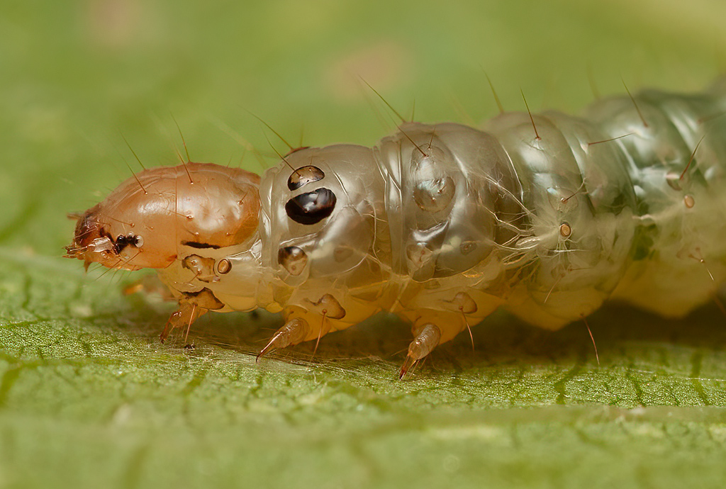
This is a dead but fresh specimen, photographed at 5X and rendered in stereo. There are some distracting bits of debris on the surface, but perhaps this at least confirms the structure of the tracheae.
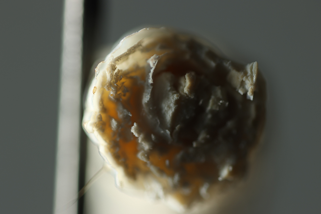
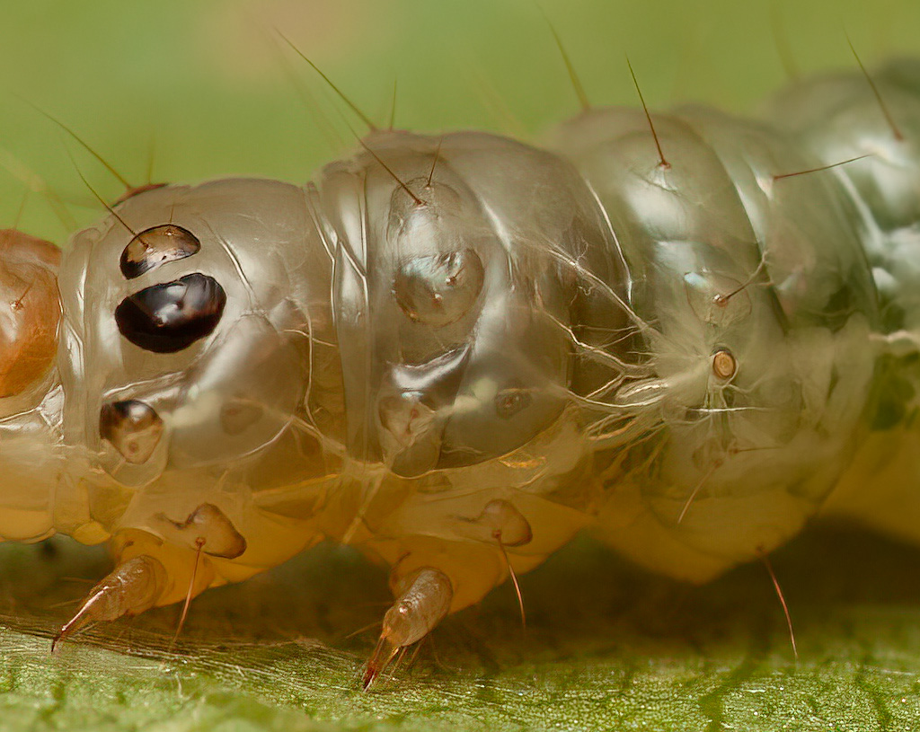
At the bottom of the preceding image, again appears that first abdominal spiracle. Here is a closer look, shot at 20X, rotated clockwise, and seriously cropped:
In the above 20X view, we can see pretty clearly that the spiracle is mostly filled up with some things that look remarkably like Q-tips. I have seen such structures before, in other caterpillar spiracles, but I have never been able to see convincingly what the structure is.
So this time I decided to push harder.
First, I spent many hours in what turned out to be a fruitless effort to photograph this spiracle with a 40X NA 0.80 apochromat. The problem is lighting -- the Q-tips are so far down inside the lip of the spiracle that by the time the available angles are limited by the high-NA objective, there's no light hitting them directly. Under those conditions, the whole scene loses detail and effectively blurs into a mass of shapes that shows no more than the 20X NA 0.42 did. Despite preparing a permanent specimen by drying in acetone, and trying several lighting schemes, I got nothing useful.
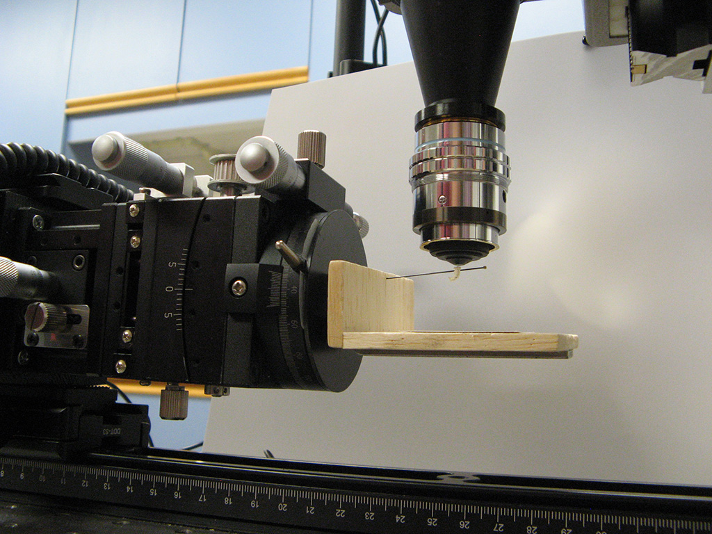
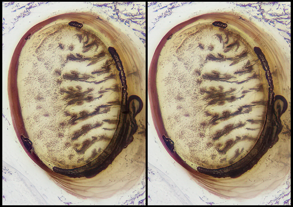
So then I switched strategies to more or less classic microscopy. I harvested another specimen (my grape vines have lots of them), killed it in boiling water, dissected the fresh specimen to extract a small piece of cuticle holding the first abdominal spiracle and essentially nothing else. Then I dried that small piece of cuticle in acetone and mounted it under a cover slip using --- wait for it --- Bondic. Yeah, Bondic. It turns out that Bondic loves chitin. It flowed nicely around and into the piece of dry cuticle, displacing essentially all the air with only a bit of manipulation. After all the air bubbles had separated from the cuticle and flowed away, I dropped a cover slip over the specimen, waited for capillary force to suck the cover slip down tight, and cured the Bondic with blue light.
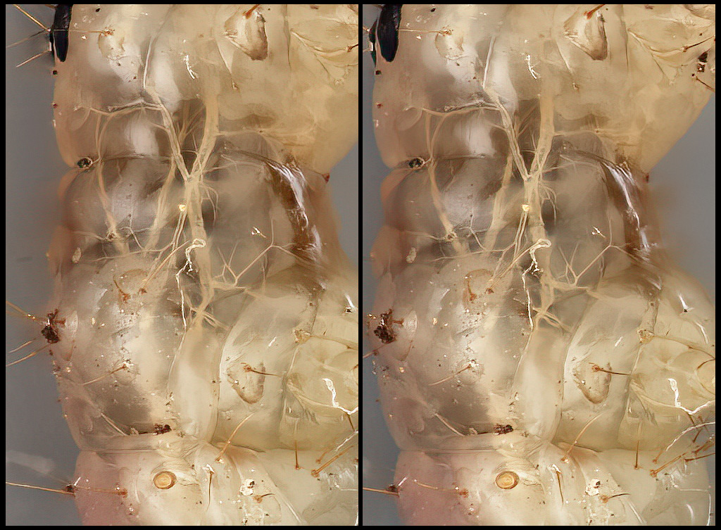
That gave me something very much like a permanent slide mount, which was simple to image with a 40X NA 0.65 bio objective with condenser illumination.
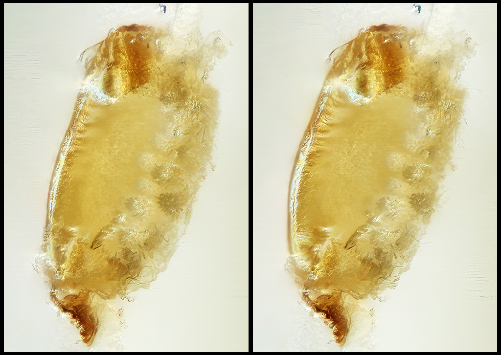
There was even enough depth left in the mount to make a decent stereo rendering. Here we have 54 frames at 0.001 mm focus step, rendered at +-6 degrees for total 12 degrees of stereo separation.
Cropped to show the whole spiracle. and very heavily sharpened to pull out as much detail as possible:
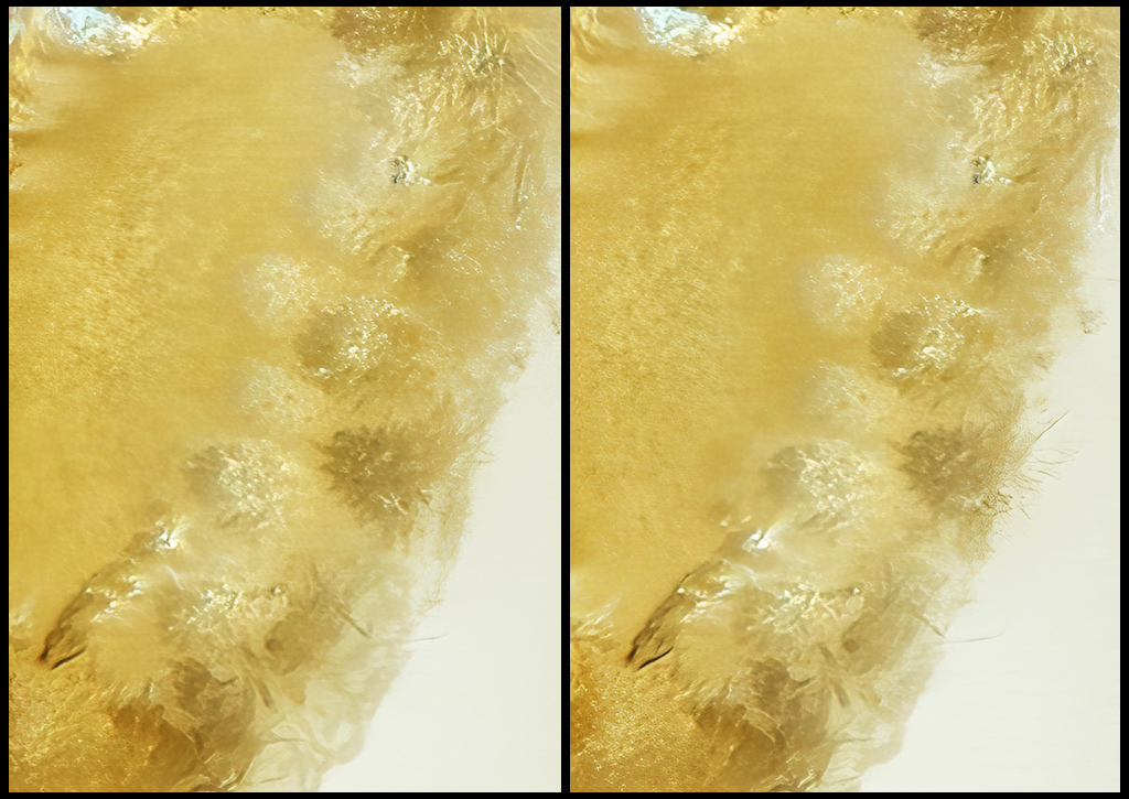
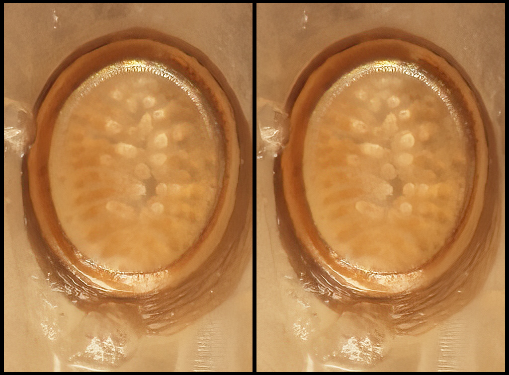
Cropped even tighter to show more structure in the hairs:
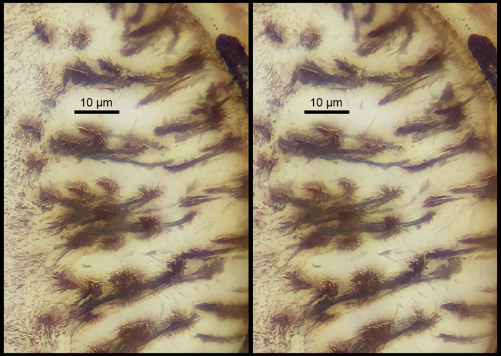
So, now I'm pretty comfortable that I understand the structure. Those "Q-tip "structures are tufts of hairs, mounted on the inner faces of the lips of the spiracle. I expect that in operation, the lips can be closed to reduce water loss, or opened to allow air exchange, but then with the air being filtered by the tufts of hairs.
It would, of course, be even more satisfying to image these things with SEM, something like HERE. But that will have to wait for some bigger budget: either time, or money, or space, or more likely all three. I am not expecting to move in that direction any time soon.
In any case, I hope you find this interesting!
--Rik