Been working on this little project for a while. I obtained a very nice Triceratium grande diatom slide, tiny pores are featured which are at times challenging to resolve and even more challenging to focus stack.
The background has been swapped with something my eye finds more pleasing, typically a circular gradient.
I will be writing a more in-depth article on my website: www.diatoms.com.au
Firstly, brightfield (60xW, PE2.5x, DMap, x2 pano):
The contrast is very low and the tiny pores are rendered at little blobs.
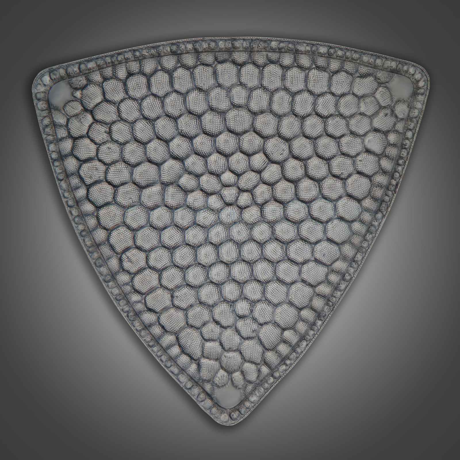
Let's spice it up a little with oblique lighting and cross polars, also known as col+pol. I am using Saul's excellent kit. Polariser used is Edmund Optic's Techspec high contrast ones. (60xW, PE2.5x, DMap, x2 pano):
Lots on contrast, somewhat 3D, this method is also known as pesudo-DIC/poor man's DIC. I would contest the second colloquialism since it has the potential to work better than DIC, especially with very thin specimens which conventional DIC struggles with.
Darkfield is challenging. My condenser only works with objectives with an NA up to 0.75. Even at 0.75, it's not great. A 40x iris objective comes to rescue. (40xO-I, PE2.5x, DMap):
I love the interference colours, but those pores are rendered as smears.
Now we're getting into established contrast methods. Phase contrast, one of my favourites, severely underrated, and quite difficult to process pictorially. (40xO-I, green interference filter, PE2.5x, DMap+PMax):
A little more work needs to be done on this one (look at the three corners). I really like it.
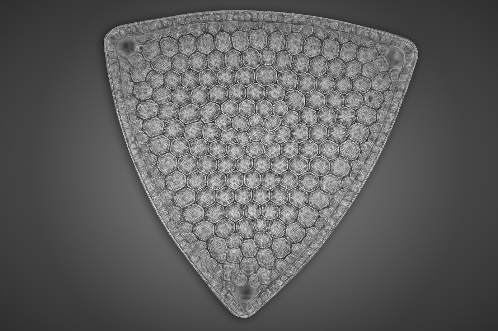
Finally, differential interference contrast (DIC, read the letters out, don't be a YouTube commenter ).
60xW, DMap:
Pesudo-3D effect is strong here, unlike Phase Contrast, the pores are perfectly rendered. Unlike COL+POL, there's minimal smears.
60xW, PE2.5x, modulated, DMap, x2 pano:
Similar to above, but with a λ plate installed. I prefer the previous one.
Hard to say which one is my favourite, any but the pure brightfield stack is great. If I had to pick one, I like Phase Contrast the most, then the first DIC, Darkfield, COL+POL, the second DIC, and brightfield. This list will probably change in a day or two.
Thanks for reading.
Microscopy method comparison: Triceratium grande Diatom
Moderators: rjlittlefield, ChrisR, Chris S., Pau
-
- Posts: 1527
- Joined: Mon Jan 15, 2018 9:23 pm
- Contact:
- rjlittlefield
- Site Admin
- Posts: 23621
- Joined: Tue Aug 01, 2006 8:34 am
- Location: Richland, Washington State, USA
- Contact:
Re: Microscopy method comparison: Triceratium grande Diatom
Really nice comparison -- thanks!
--Rik
--Rik
-
- Posts: 5090
- Joined: Sun Jan 15, 2012 12:31 pm
Re: Microscopy method comparison: Triceratium grande Diatom
Interesting series. Especially liked the last two.
Mike
Mike
Michael Reese Much FRMS EMS Bethlehem, Pennsylvania, USA
Re: Microscopy method comparison: Triceratium grande Diatom
Yes, really interesting comparison, and fantastic results even if you had only posted one of them. I'll probably be alone in this opinion but I find that I like the simple first one the best.
-
- Posts: 1527
- Joined: Mon Jan 15, 2018 9:23 pm
- Contact:
Re: Microscopy method comparison: Triceratium grande Diatom
Thanks for the comments!
It's all a matter of taste with these, some like the glassy reflected light look too. I don't have such a slide yet, simply putting a piece of black cardboard under the slide doesn't work with these. Would like to add Hoffman Modulation Contrast and some other more exotic contrast methods in the future.
Re: Microscopy method comparison: Triceratium grande Diatom
Thanks for this. These comparisons are really interesting and have inspired me to try my own (minus the DIC which is beyond my means). Could you update this thread when your article is published?
Mike
Mike
-
- Posts: 1527
- Joined: Mon Jan 15, 2018 9:23 pm
- Contact:
Re: Microscopy method comparison: Triceratium grande Diatom
Sure, it would be too long, likely very similar to this post. I only recently purchased the domain name and I'm gonna try and see which webpage builder to go with. Currently, it just redirect to my old wordpress blog.
Re: Microscopy method comparison: Triceratium grande Diatom
I like the detail, contrast, and color of the 5th one. They are all very nice though!
Re: Microscopy method comparison: Triceratium grande Diatom
Macro_Cosmos,
Haven't you tried your Leitz Heine in darkfield mode? With that you can get darkfield in objective NA of 1.1, depending on diatom (thinner ones with cleaner background respond better).
Super thin diatoms, such as Amphipleura pellucida, surely "dot" better under oblique (COL) + pol than DIC.
With most diatoms though, I find Heine darkfield or DIC images more pleasing, than oblique (I dislike smearing or shadow effects) or phase contrast.
-
- Posts: 1527
- Joined: Mon Jan 15, 2018 9:23 pm
- Contact:
Re: Microscopy method comparison: Triceratium grande Diatom
My Heine condenser is still in transit, it's been a good 10 days... no luck getting it before May, not going to hold my breath for it. I paid 70 euros for the "premium shipping" add-on... I expected a week.zzffnn wrote: ↑Sun Apr 25, 2021 5:24 pmMacro_Cosmos,
Haven't you tried your Leitz Heine in darkfield mode? With that you can get darkfield in objective NA of 1.1, depending on diatom (thinner ones with cleaner background respond better).
Super thin diatoms, such as Amphipleura pellucida, surely "dot" better under oblique (COL) + pol than DIC.
With most diatoms though, I find Heine darkfield or DIC images more pleasing, than oblique (I dislike smearing or shadow effects) or phase contrast.
You're right, with very thin specimens, DIC doesn't work well unless one has access to the "HC = high contrast" types, not me. COL is usually very good, phase contrast from my experience works very well. The same applies for very thick specimens where COL is nice, DIC can produce false results or weird affects (need "HR = high resolution" for >200um specimens), just brightfield is nice too.